. 2021 Feb:64:103196.
doi: 10.1016/j.ebiom.2020.103196. Epub 2021 Jan 19.

Repopulation of decellularised articular cartilage by laser-based matrix engraving

Affiliations

Repopulation of decellularised articular cartilage by laser-based matrix engraving

S Nürnberger et al. EBioMedicine. 2021 Feb.

Abstract

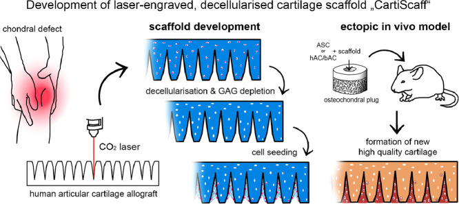
Background: In spite of advances in the treatment of cartilage defects using cell and scaffold-based therapeutic strategies, the long-term outcome is still not satisfying since clinical scores decline years after treatment. Scaffold materials currently used in clinical settings have shown limitations in providing suitable biomechanical properties and an authentic and protective environment for regenerative cells. To tackle this problem, we developed a scaffold material based on decellularised human articular cartilage.

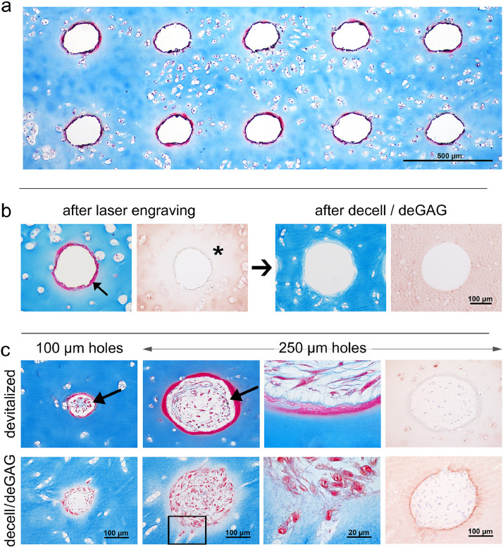
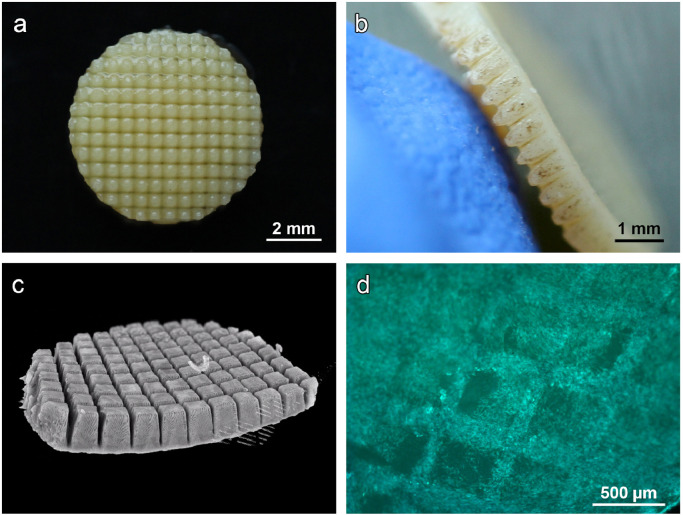
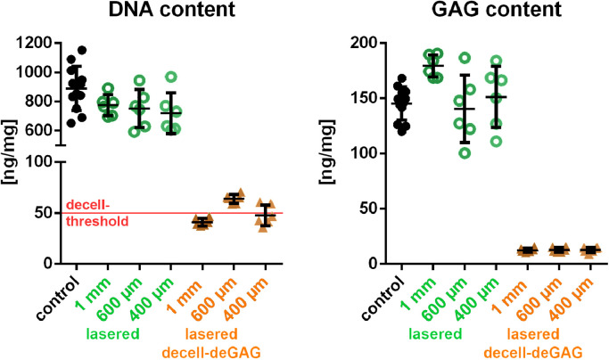
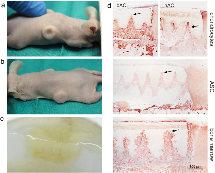
Methods: Human articular cartilage matrix was engraved using a CO2 laser and treated for decellularisation and glycosaminoglycan removal. Characterisation of the resulting scaffold was performed via mechanical testing, DNA and GAG quantification and in vitro cultivation with adipose-derived stromal cells (ASC). Cell vitality, adhesion and chondrogenic differentiation were assessed. An ectopic, unloaded mouse model was used for the assessment of the in vivo performance of the scaffold in combination with ASC and human as well as bovine chondrocytes. The novel scaffold was compared to a commercial collagen type I/III scaffold.

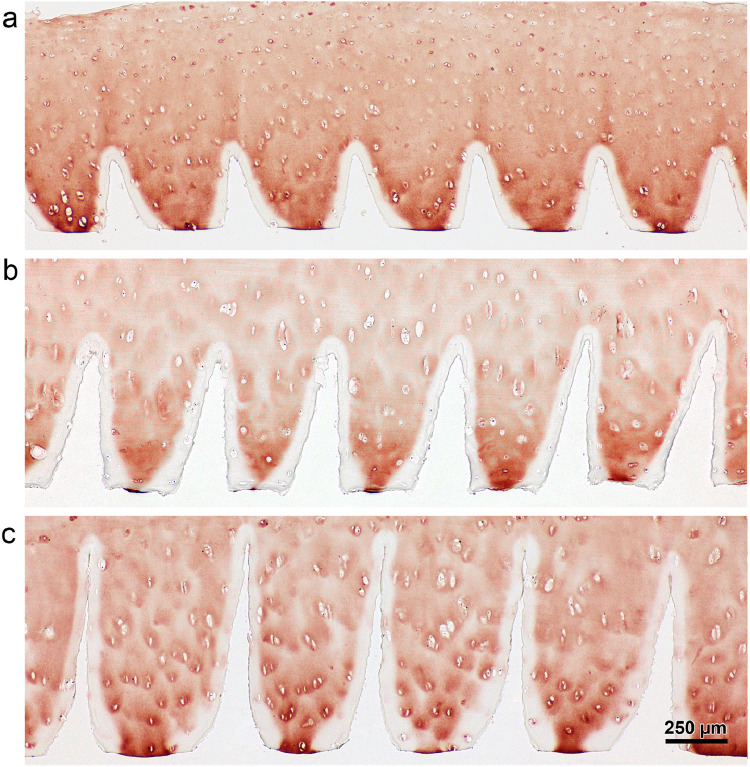
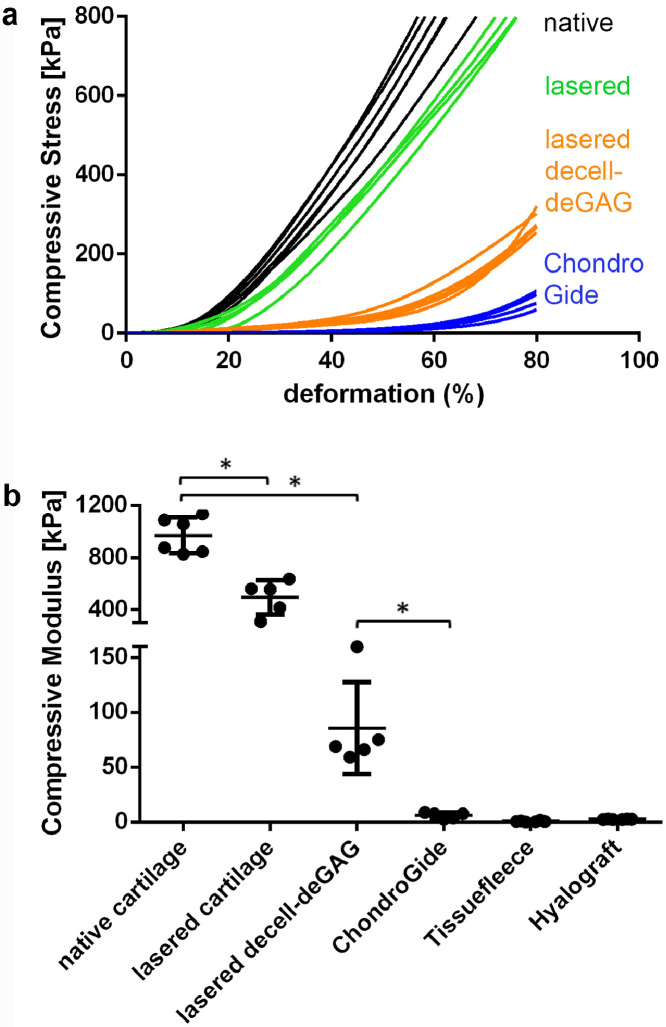
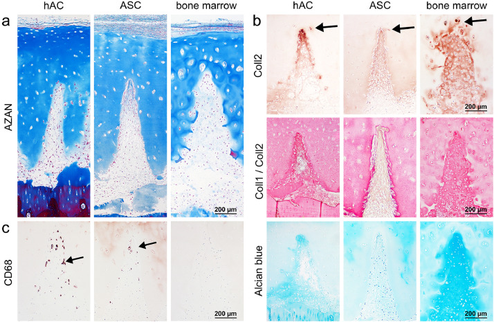
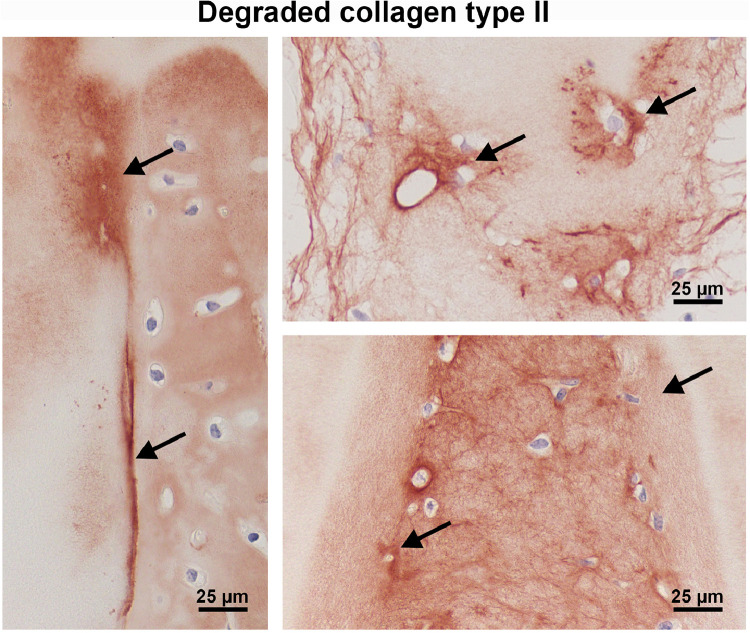
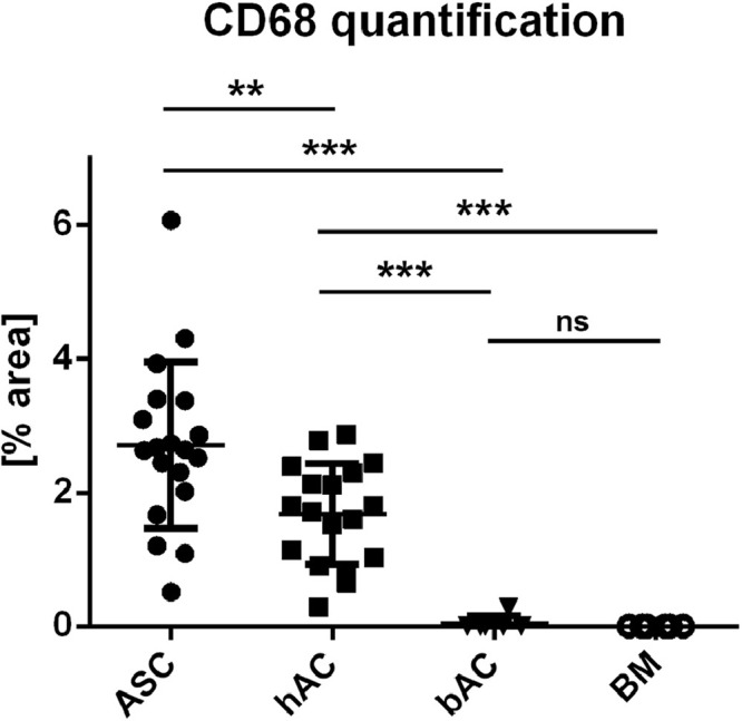
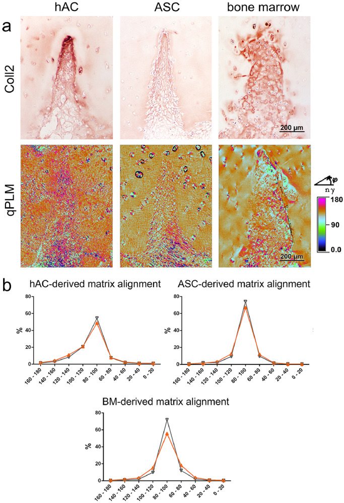
Findings: Crossed line engravings of the matrix allowed for a most regular and ubiquitous distribution of cells and chemical as well as enzymatic matrix treatment was performed to increase cell adhesion. The biomechanical characteristics of this novel scaffold that we term CartiScaff were found to be superior to those of commercially available materials. Neo-tissue was integrated excellently into the scaffold matrix and new collagen fibres were guided by the laser incisions towards a vertical alignment, a typical feature of native cartilage important for nutrition and biomechanics. In an ectopic, unloaded in vivo model, chondrocytes and mesenchymal stromal cells differentiated within the incisions despite the lack of growth factors and load, indicating a strong chondrogenic microenvironment within the scaffold incisions. Cells, most noticeably bone marrow-derived cells, were able to repopulate the empty chondrocyte lacunae inside the scaffold matrix.

Interpretation: Due to the better load-bearing, its chondrogenic effect and the ability to guide matrix-deposition, CartiScaff is a promising biomaterial to accelerate rehabilitation and to improve long term clinical success of cartilage defect treatment.

Funding: Austrian Research Promotion Agency FFG ("CartiScaff" #842455), Lorenz Böhler Fonds (16/13), City of Vienna Competence Team Project Signaltissue (MA23, #18-08).

Keywords: Cartilage regeneration; Decellularisation; Ectopic animal model; Laser engraving; Mechanical testing; Repopulation.


Conflict of interest statement

Declaration of Competing Interests Dr. Grillari reports that he is Co-founder and shareholder of Evercyte GmbH, the company providing the ASC/TERT1 cells used in this study. Dr. Nürnberger reports grants from Austrian Research Promotion Agency FFG, grants from Lorenz Böhler Society, during the conduct of the study; In addition, Dr. Nürnberger and Dr. Redl have a patent WO 2018220047 A1 issued. Dr. Redl CEO of Trauma Care Consult. Dr. Heimel and Mrs. Schneider report grants from Austrian Research Promotion Agency FFG grants from Lorenz Böhler Society, during the conduct of the study. Dr. Wolbank reports grants from Österreichische Forschungsförderungsgesellschaft, during the conduct of the study.

Figures

Image, graphical abstract
Graphical abstract
Fig 1
Fig. 1
Laser-engraved holes: effects on matrix and reseeding. a) Laser engraving enables the generation of highly regular hole patterns. b) The laser process leads to matrix densification along the edge visible as red ring on AZAN staining (arrow) and altered collagen type II immunoreactivity surrounding the holes (asterisk). Both effects disappeared after decell-deGAG treatment, even though leaving the collagen staining slightly less intense than untreated, unlasered native cartilage (n > 3). c) All types of holes (100 µm, 250 µm diameter, devitalized, decell-deGAG) are successfully repopulated, yet on devitalized samples cells are in a distance to the densified scaffold surface (arrows) with few fibrous connections (magnification); on decell-deGAG samples repopulation begins at the scaffold border and cells invade accessible lacunae (square and magnification thereof); n = 3.
Fig 2
Fig. 2
Laser-engraved line pattern: direct relation between increasing laser runs and increasing incision depth. Collagen type II stained cross-sections of cartilage tissue subjected to laser engraving a) 3 runs, b) 6 runs and c) 9 runs. There is hardly any effect on the incision width on surface level or the amount of collagen type II denaturation. n > 3.
Fig 3
Fig. 3
Laser-engraved crossed line incisions: spatial appearance. a) Macroscopic top view and b) side view of cartilage biopsy with laser-engraved cross-wise line incisions and c) µCT giving a three-dimensional impression of the precise and highly regular grid pattern. d) Calcein-AM staining of ASC seeded on laser-engraved cartilage disc showing a high density of vital cells inside the incisions.
Fig 4
Fig. 4
DNA and GAG quantification of the laser-engraved, decellularised and GAG-deprived cartilage matrix. DNA content is reduced from 889+151 to 51+/−6 ng/mg dry weight: to 48+/−10 ng/mg in 400 µm thick samples, 64+/−4.4 ng/mg in 600 µm thick samples to 41 +/−3.8 in 1 mm thick samples. GAG content is lowered from 145+/−15 to 13+2.0 µg/mg dry weight. Red line at 50 ng/mg in the DNA control marks the threshold of decellularisation. control n = 12; lasered and lasered decell-deGAG samples n=6. (For interpretation of the references to colour in this figure legend, the reader is referred to the web version of this article.)
Fig 5
Fig. 5
Influence of engraved grid patterns and final scaffold pretreatment on compressive stiffness and modulus. a) Native cartilage reaches 61 +/−4 % and lasered cartilage 74 +/−2 % deformation at the maximal load of 800 kPa while decell-deGAG scaffolds is deformed 80% with 284 +/−26 kPa. 80% ChondroGide-deformation is already reached with 88 +/−20 kPa. b) At 17-20% deformation, compressive modulus of cartilage with laser-engraved grid patterns is reduced to 51% compared to native cartilage. Laser-engraved decell-deGAG cartilage retains 6–16% of its native compressive modulus, leaving it 10 to 26 fold stronger than the commercially available controls; data shown as mean + SD. * P = 0.05, n = ≥5 (Mann-Whitney-U-Tests).
Fig 6
Fig. 6
In vitro seeding tests on cartilage with laser-engraved grid patterns. a) AZAN staining and b) µCT imaging of ASC on laser-engraved cartilage scaffolds after one week of cultivation in proliferation medium followed by two weeks of low dose differentiation medium. On devitalised samples (left) a gap remains between de-novo matrix and the scaffold surface (arrow), while on decell-deGAG treated samples (right) cells adhere tightly to the scaffold matrix. c) After five weeks cultivation of ASC on decell-deGAG treated scaffolds in differentiation medium, (immuno-)histochemistry reveals the presence of neo-cartilage with a dense matrix staining for collagen type II and GAG (Alcian blue) and fully integrated with the scaffold matrix. Polarization light microscopy of picro sirius red stained samples shows collagen fibres aligned along the cutting edges and perpendicular to the cartilage surface (insert). Representative consecutive sections; n = 3. (For interpretation of the references to colour in this figure legend, the reader is referred to the web version of this article.)
Fig 7
Fig. 7
Performance of reseeded scaffolds in an osteochondral defect model in vivo. Macroscopic images reveal that after six weeks the experimental defects a) appear to be empty in the control group, while b) defects filled with reseeded scaffolds feature a smooth surface. c) Cross-section through the osteochondral plug proving the filling of the defect with scaffold and new matrix. d) The histological overview sections show the scaffolds and neo-tissue within the defects and differences in distribution of collagen type II. While neo-tissue derived from bovine chondrocytes (bAC) stains regular and intensely, human chondrocytes (hAC) or ASC/TERT1-derived matrix stains strongest within the incisions (tip effect). Bone marrow invading from the subchondral space of the plug forms an intensively stained hyaline tissue all over the defect. The incision edges are more irregular than when chondrocytes or ASC/TERT1 fill the incisions. At some sites collagen type II producing cells are visible inside the scaffold close to the incision edge (arrows). n = 6.
Fig 8
Fig. 8
Chondrocytes (hAC), ASC/TERT1 or bone marrow cells in the incisions of decell-deGAG treated scaffolds with laser-engraved grid patterns in vivo. (Immuno-)Histochemistry shows a a) regular distribution of cells inside the incisions and b) newly synthesized matrix. Human chondrocyte-derived matrix displays a gradient of increasing staining intensity of collagen type II (Coll2=brown in single-staining) within the incisions (tip effect) and a dominance of collagen type II staining over collagen type I in the double staining (Coll2=pink, Coll1=brown in double-staining). A gradient towards the tip is also visible for GAG in Alcian blue staining. In the ASC/TERT1-seeded scaffolds collagen type II is found along the whole incision with slightly increasing density towards the tip but still generally weaker staining in comparison to the chondrocyte group. Also in the double staining collagen type I is more dominant (brown instead of red in Coll1/Coll2 double staining) and the GAG-staining faint (Alcian blue). Matrix derived from invaded bone marrow cells stains regularly and strong for collagen type II without any visible collagen type I staining (only pink Coll1/Coll2 double staining) and strong GAG (Alcian blue) staining. In all groups some cells invaded empty lacunae inside the scaffold matrix (arrows). c) Staining of CD68 positive cells (arrows) indicates the presence of macrophages in the ASC/TERT1 and hAC group but not in the dense, bone-marrow involving neo-cartilage. n = 6. (For interpretation of the references to colour in this figure legend, the reader is referred to the web version of this article.)
Fig 9
Fig. 9
Localisation of degraded collagen type II (CTX-II; arrows) at the edge of the scaffold or in the circumference of invading cells. Incisions of CartiScaff implanted in osteochondral defects with bone marrow (left), hAC (top) and bAC (below) and kept for six weeks in vivo.
Fig 10
Fig. 10
Macrophage quantification via CD68 positive areas within the incisions. Most staining is present in the ASC/TERT1 (ASC) group followed by the hAC group. In bAC only one and in the bone marrow group no incision contains positive areas. data shown as mean + SD. ** P = 0,0036 *** P < 0,0001 (Mann-Whitney U test) ASC/TERT1 n = 19; hAC n= 18; bAC n= 6; BM n= 6.
Fig 11
Fig. 11
Collagen fibre alignment inside the neo-cartilage. a) Collagen type II staining (same sites as in Fig. 8B) and the corresponding qPLM images, with the fibre orientation for each pixel (0.5 µm) assigned to a colour range. b) Quantitative distribution of fibre orientation reveals relatively regular fibres alignment inside the engraved incisions (orange curve), corresponding to the alignment inside the scaffold (black curve), both with the majority of fibres arranged in an angle of 80° to 100° to the scaffold surface. BM = bone marrow. (For interpretation of the references to colour in this figure legend, the reader is referred to the web version of this article.)

References

    1. Marlovits S., Zeller P., Singer P., Resinger C., Vécsei V. Cartilage repair: generations of autologous chondrocyte transplantation. Eur J Radiol. 2006;57:24–31. doi: 10.1016/j.ejrad.2005.08.009. - DOI - PubMed
    1. Brittberg M. Cell carriers as the next generation of cell therapy for cartilage repair: a review of the matrix-induced autologous chondrocyte implantation procedure. Am J Sports Med. 2010;38:1259–1271. doi: 10.1177/0363546509346395. - DOI - PubMed
    1. Cherubino P., Grassi F.A., Bulgheroni P., Ronga M. Autologous chondrocyte implantation using a bilayer collagen membrane: a preliminary report. J Orthop Surg (Hong Kong) 2003;11(1):10–15. doi: 10.1177/230949900301100104. - DOI - PubMed
    1. Aldrian S., Zak L., Wondrasch B., Albrecht C., Stelzeneder B., Binder H., Kovar F., Trattnig S., Marlovits S. Clinical and radiological long-term outcomes after matrix-induced autologous chondrocyte transplantation: a prospective follow-up at a minimum of 10 years. Am J Sports Med. 2014;42(11):2680–2688. doi: 10.1177/0363546514548160. - DOI - PubMed
    1. Zak L., Albrecht C., Wondrasch B., Widhalm H., Vekszler G., Trattnig S., Marlovits S., Aldrian S. Results 2 years after matrix-associated autologous chondrocyte transplantation using the novocart 3D scaffold: an analysis of clinical and radiological data. Am J Sports Med. 2014;42(7):1618–1627. doi: 10.1177/0363546514532337. - DOI - PubMed