. 2021 Mar 11;87(7):e02875-20.
doi: 10.1128/AEM.02875-20. Print 2021 Mar 11.

Inactivation of GalU Leads to a Cell Wall-Associated Polysaccharide Defect That Reduces the Susceptibility of Enterococcus faecalis to Bacteriolytic Agents

Affiliations

Inactivation of GalU Leads to a Cell Wall-Associated Polysaccharide Defect That Reduces the Susceptibility of Enterococcus faecalis to Bacteriolytic Agents

Jun Kurushima et al. Appl Environ Microbiol. .

Abstract

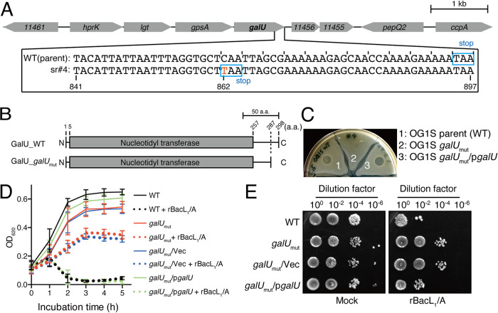
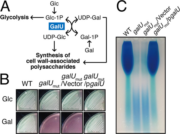
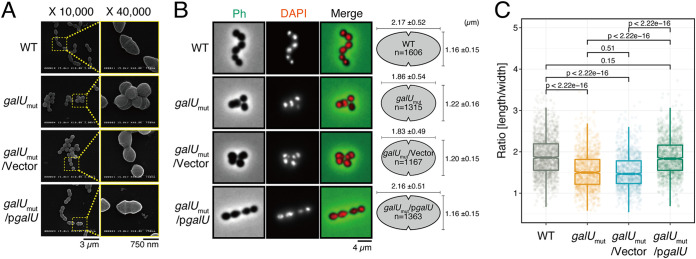
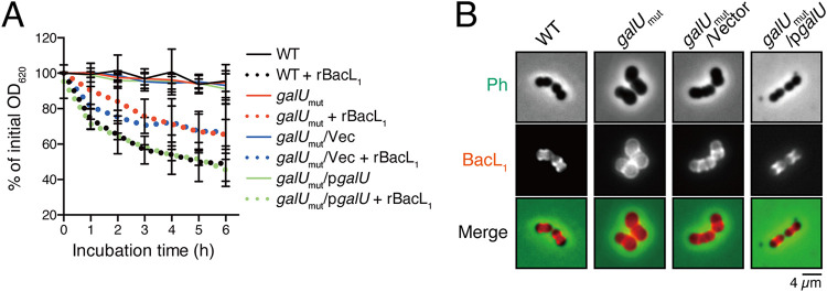
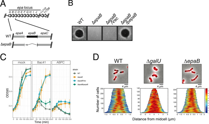
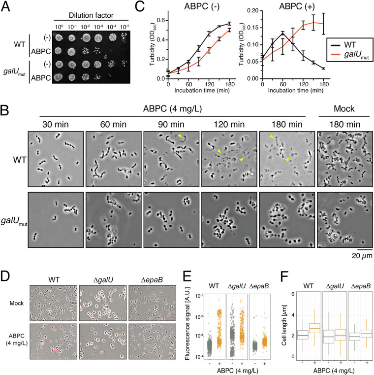
Enterococcal plasmid-encoded bacteriolysin Bac41 is a selective antimicrobial system that is considered to provide a competitive advantage to Enterococcus faecalis cells that carry the Bac41-coding plasmid. The Bac41 effector consists of the secreted proteins BacL1 and BacA, which attack the cell wall of the target E. faecalis cell to induce bacteriolysis. Here, we demonstrated that galU, which encodes UTP-glucose-1-phosphate uridylyltransferase, is involved in susceptibility to the Bac41 system in E. faecalis Spontaneous mutants that developed resistance to the antimicrobial effects of BacL1 and BacA were revealed to carry a truncation deletion of the C-terminal amino acid (aa) region 288 to 298 of the translated GalU protein. This truncation resulted in the depletion of UDP-glucose, leading to a failure to utilize galactose and produce the enterococcal polysaccharide antigen (EPA), which is expressed abundantly on the cell surface of E. faecalis This cell surface composition defect that resulted from galU or EPA-specific genes caused an abnormal cell morphology, with impaired polarity during cell division and alterations of the limited localization of BacL1 Interestingly, these mutants had reduced susceptibility to beta-lactams besides Bac41, despite their increased susceptibility to other bacteriostatic antimicrobial agents and chemical detergents. These data suggest that a complex mechanism of action underlies lytic killing, as exogenous bacteriolysis induced by lytic bacteriocins or beta-lactams requires an intact cell physiology in E. faecalisIMPORTANCE Cell wall-associated polysaccharides of bacteria are involved in various physiological characteristics. Recent studies demonstrated that the cell wall-associated polysaccharide of Enterococcus faecalis is required for susceptibility to bactericidal antibiotic agents. Here, we demonstrated that a galU mutation resulted in resistance to the enterococcal lytic bacteriocin Bac41. The galU homologue is reported to be essential for the biosynthesis of species-specific cell wall-associated polysaccharides in other Firmicutes In E. faecalis, the galU mutant lost the E. faecalis-specific cell wall-associated polysaccharide EPA (enterococcal polysaccharide antigen). The mutant also displayed reduced susceptibility to antibacterial agents and an abnormal cell morphology. We demonstrated here that galU was essential for EPA biosynthesis in E. faecalis, and EPA production might underlie susceptibility to lytic bacteriocin and antibiotic agents by undefined mechanisms.

Keywords: Enterococcus; Enterococcus faecalis; UDP-glucose phosphorylase; antibiotic resistance; bacteriocin; beta-lactam; beta-lactams; cell death; cell morphology; galactose; imaging; morphogenesis; polysaccharide.


Figures

FIG 1
FIG 1
Genetic mutation found in the strain spontaneously resistant to the antimicrobial activity of Bac41. (A) Nucleotide sequence alignment of the galU (OG1RF_11457; Gene ID 12287400) genes of the E. faecalis OG1S parent strain (upper) and the OG1S isogenic mutant strain (sr#4) that spontaneously acquired resistance to the antimicrobial activity of Bac41 (lower). Scale bar, 1 kb. (B) Amino acid sequence alignment of the translation products of the galU genes of the E. faecalis OG1S parent strain (upper) and the OG1S isogenic mutant strain (lower). Scale bar, 50 aa. (C) A mixture of recombinant BacL1-His and BacA-His proteins (25 ng each) was spotted onto THB soft agar (0.75%) containing the indicator strain E. faecalis OG1S (parent, 1), the spontaneously resistant strain with the galU mutation (galUmut, 2), and the galUmut complemented strain via the transexpression of wild-type GalU from the pgalU plasmid (galUmut/pgalU, 3). The plate was incubated at 37°C for 24 h, and the formation of halos was evaluated. (D) Overnight cultures of E. faecalis strains, including OG1S (WT), the galU mutant (galUmut), the vector control strain of galUmut (galUmut/Vec), and the complemented galUmut strain (galUmut/pgalU), were inoculated into fresh THB broth at a 5-fold dilution. A mixture of recombinant BacL1-His (5 μg/ml) and BacA-His (5 μg/ml) was added to the bacterial suspension and incubated at 37°C. The turbidity was monitored during the incubation period. The data for each case are presented as the means ± standard deviations (SD) (error bars) from three independent experiments. (E) The E. faecalis strains were treated with rBacL1 and rBacA (5 μg/ml each) at 37°C for 4 h, as described for panel D. The bacterial suspensions were serially diluted 100-fold with fresh THB and then spotted onto a THB agar plate, followed by incubation overnight. Colony formation was evaluated as a measure of bacterial viability.
FIG 2
FIG 2
galU is essential for galactose fermentation and cell surface polysaccharide production. (A) The Leloir pathway of galactose metabolism is illustrated. GalU generates UDP-glucose (UDP-Glc) from glucose-1-phosphate (Glc-1P) and ATP. (B) The E. faecalis wild-type, galUmut, galUmut/Vec, and galUmut/pgalU strains were grown in HI agar medium supplemented with phenol red as a pH indicator and glucose (Glc) or galactose (Gal) as the fermentation source. (C) The E. faecalis wild-type, galUmut, galUmut/Vec, and galUmut/pgalU strains were grown in THB broth supplemented with glucose, and the cell wall-associated polysaccharides were prepared as described in Materials and Methods. The resulting polysaccharides were separated via 10% acrylamide gel electrophoresis, followed by staining with the Stains-All reagent.
FIG 3
FIG 3
galU is essential for maintaining cell morphology. (A) The E. faecalis wild-type, galUmut, galUmut/Vec, and galUmut/pgalU strains were grown on cover glasses, followed by chemical fixation. The samples were subjected to osmium coating and analyzed under the scanning electron microscope. Scale bars, 3 μm (left) and 750 nm (right). (B) Overnight cultures of the E. faecalis wild-type, galUmut, galUmut/Vec, and galUmut/pgalU strains were diluted 5-fold with fresh THB broth and incubated, chemically fixed, and mounted with DAPI for DNA visualization (red), followed by analysis via fluorescence microscopy. Phase contrast (Ph) is pseudocolored (green) in the merged image. Scale bar, 4 μm. The panel on the right represents mean ± SD individual cell length and width; at least >1,000 cells were counted for each strain. (C) Cellular length/width ratio of single cells, calculated based on phase-contrast images in panel B, were plotted. t test was used for statistical analysis.
FIG 4
FIG 4
galU inactivation alters the interaction of BacL1 with the cell wall of E. faecalis. (A) Cell wall fractions prepared from E. faecalis wild-type (black), galUmut (red), galUmut/Vec (blue), and galUmut/pgalU (green) strains in exponential phase were diluted with PBS. Recombinant BacL1 (rBacL1; 5 μg/ml, dotted lines) was added to the cell wall suspension, and the mixture was incubated at 37°C. The turbidity at 600 nm was quantified at the indicated times during incubation. The presented values are the percentages of the initial turbidity for the respective samples. The PBS-treated sample (mock) is presented as a negative control (straight lines). The data are presented as the means ± SD (error bars) from four independent experiments. (B) Overnight cultures of the E. faecalis wild-type, galUmut, galUmut/Vec, and galUmut/pgalU strains were diluted 5-fold with fresh THB broth and incubated with HiLyte Fluor 555 fluorescent dye-labeled (red) BacL1 (5 μg/ml), followed by analysis via fluorescence microscopy. Phase contrast (Ph) is pseudocolored (green) in the merged images. Scale bar, 4 μm.
FIG 5
FIG 5
Effect of epaB deletion on the binding affinity of BacL1 and susceptibility to Bac41 activity. (A) Genomic organization of the epa locus in E. faecalis OG1RF and construction of an epaB deletion mutant. (B) A mixture of the recombinant BacL1-His and BacA-His proteins (25 ng each) was spotted onto THB soft agar (0.75%) containing the E. faecalis wild-type indicator strain and ΔepaB, ΔepaB/Vec, and ΔepaB/pepaB strains. The plate was incubated at 37°C for 24 h, and the formation of halos was evaluated. (C) The overnight cultures of the E. faecalis OG1RF (WT), ΔepaB, ΔepaB/Vec, and ΔepaB/pepaB strains were inoculated into fresh THB broth at a 10-fold dilution in the presence or absence of ABPC (4 mg/liter) or Bac41 (BacL1 and BacA, 2.5 μg/ml each), followed by incubation at 37°C. The turbidity (OD595) was monitored during the incubation period. The data for each case are presented as the percentage of the initial turbidity. The data are presented as the means ± SD (error bars) from three independent experiments. (D) Overnight cultures of the E. faecalis wild-type, ΔgalU, and ΔepaB strains were diluted 5-fold with fresh THB broth and incubated with HiLyte Fluor 555 fluorescent dye-labeled (red) BacL1_SH3-His (5 μg/ml), followed by analysis via fluorescence microscopy.
FIG 6
FIG 6
Different bacteriolysis phenotypes induced by ampicillin treatment (ABPC) in the E. faecalis wild-type strain or the galUmut mutant. (A) The E. faecalis strains OG1S (WT) and the galUmut mutant were treated with ABPC (4 mg/liter) at 37°C for 3 h. The bacterial suspensions were serially diluted 10-fold with fresh THB and then spotted onto a THB agar plate, followed by incubation overnight. Colony formation was evaluated as a measure of bacterial viability. (B) Confluent cultures of the E. faecalis OG1S (WT) and galUmut strains were diluted 10-fold with fresh THB containing ABPC (4 mg/liter), followed by incubation at 37°C. The bacterial suspension was mounted on a slide and analyzed via microscopy (phase contrast) at the indicated time points: 30, 60, 90, 120, and 180 min after treatment. The yellow arrowheads indicate the cell debris generated by ABPC-induced bacteriolysis. Cells incubated under identical conditions except for the absence of ABPC (Mock) are represented in the right panel as a reference. Scale bar, 20 μm. (C) The overnight cultures of the E. faecalis OG1S (WT) and galUmut strains were inoculated into fresh THB broth at a 10-fold dilution in the presence or absence of ABPC (4 mg/liter), followed by incubation at 37°C. The turbidity was monitored during the incubation period. The data for each case are presented as the percentage of the initial turbidity. The data are presented as the means ± SD (error bars) from three independent experiments. (D) The overnight cultures of the E. faecalis OG1RF (WT), ΔgalU, and ΔepaB strains were inoculated into fresh THB broth at a 10-fold dilution in the presence or absence of ABPC (4 mg/liter), followed by incubation at 37°C for 1 h with EthD (2 mg/liter). The bacterial suspension was mounted onto a slide and analyzed via fluorescence microscopy. Images are shown as merges of phase contrast and red fluorescence signals (EthD). (E and F) Quantification of population with red fluorescence intensity (E) and cell length (F) on each cell was generated based on data from panel D. A.U., arbitrary units.

Similar articles

Cited by

References

    1. Arias CA, Murray BE. 2012. The rise of the Enterococcus: beyond vancomycin resistance. Nat Rev Microbiol 10:266–278. doi:10.1038/nrmicro2761. - DOI - PMC - PubMed
    1. Nes IF, Diep DB, Holo H. 2007. Bacteriocin diversity in Streptococcus and Enterococcus. J Bacteriol 189:1189–1198. doi:10.1128/JB.01254-06. - DOI - PMC - PubMed
    1. Nes IF, Clewell DB, Ike Y, Shankar N, Diep DB. 2014. Enterococcal bacteriocins and antimicrobial proteins that contribute to niche control. In Gilmore MS, Clewell DB, Ike Y, et al.. (ed), Enterococci: from commensals to leading causes of drug resistant infection. Massachusetts Eye and Ear Infirmary, Boston, MA. - PubMed
    1. Jack RW, Tagg JR, Ray B. 1995. Bacteriocins of gram-positive bacteria. Microbiol Rev 59:171–200. doi:10.1128/MMBR.59.2.171-200.1995. - DOI - PMC - PubMed
    1. Field D, Hill C, Cotter PD, Ross RP. 2010. The dawning of a “golden era” in lantibiotic bioengineering. Mol Microbiol 78:1077–1087. doi:10.1111/j.1365-2958.2010.07406.x. - DOI - PubMed

Publication types

MeSH terms

LinkOut - more resources