. 2021 Mar 10;41(10):2264-2273.
doi: 10.1523/JNEUROSCI.1871-20.2020. Epub 2021 Jan 22.

α-Synuclein Oligomers Induce Glutamate Release from Astrocytes and Excessive Extrasynaptic NMDAR Activity in Neurons, Thus Contributing to Synapse Loss

Affiliations

α-Synuclein Oligomers Induce Glutamate Release from Astrocytes and Excessive Extrasynaptic NMDAR Activity in Neurons, Thus Contributing to Synapse Loss

Dorit Trudler et al. J Neurosci. .

Abstract

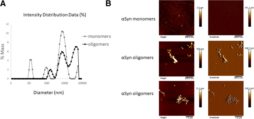
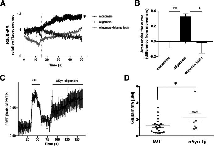
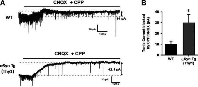
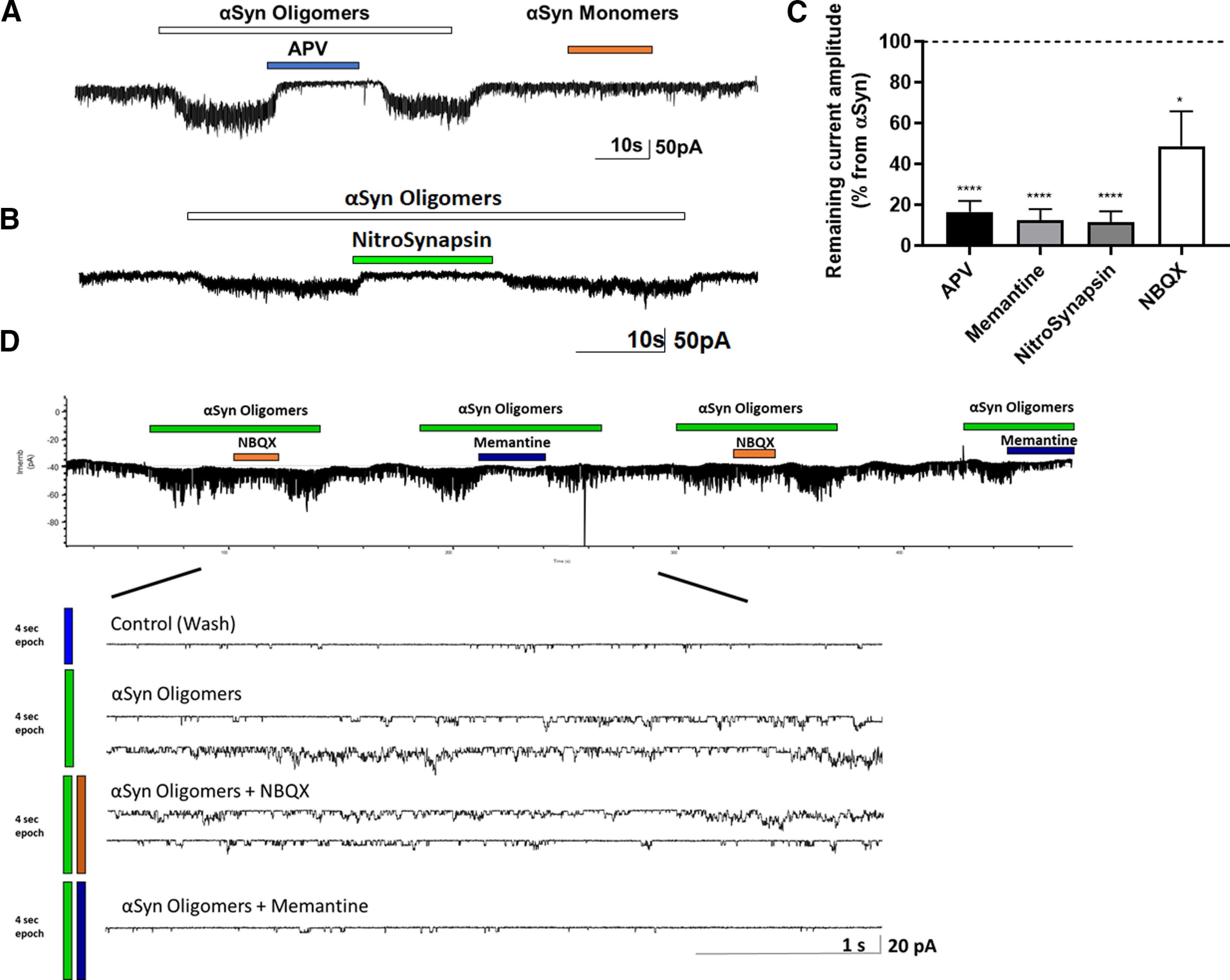
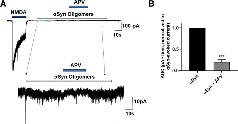
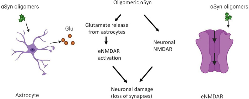
Synaptic and neuronal loss are major neuropathological characteristics of Parkinson's disease. Misfolded protein aggregates in the form of Lewy bodies, comprised mainly of α-synuclein (αSyn), are associated with disease progression, and have also been linked to other neurodegenerative diseases, including Lewy body dementia, Alzheimer's disease, and frontotemporal dementia. However, the effects of αSyn and its mechanism of synaptic damage remain incompletely understood. Here, we show that αSyn oligomers induce Ca2+-dependent release of glutamate from astrocytes obtained from male and female mice, and that mice overexpressing αSyn manifest increased tonic release of glutamate in vivo In turn, this extracellular glutamate activates glutamate receptors, including extrasynaptic NMDARs (eNMDARs), on neurons both in culture and in hippocampal slices of αSyn-overexpressing mice. Additionally, in patch-clamp recording from outside-out patches, we found that oligomerized αSyn can directly activate eNMDARs. In organotypic slices, oligomeric αSyn induces eNMDAR-mediated synaptic loss, which can be reversed by the drug NitroSynapsin. When we expose human induced pluripotent stem cell-derived cerebrocortical neurons to αSyn, we find similar effects. Importantly, the improved NMDAR antagonist NitroSynapsin, which selectively inhibits extrasynaptic over physiological synaptic NMDAR activity, protects synapses from oligomeric αSyn-induced damage in our model systems, thus meriting further study for its therapeutic potential.SIGNIFICANCE STATEMENT Loss of synaptic function and ensuing neuronal loss are associated with disease progression in Parkinson's disease (PD), Lewy body dementia (LBD), and other neurodegenerative diseases. However, the mechanism of synaptic damage remains incompletely understood. α-Synuclein (αSyn) misfolds in PD/LBD, forming Lewy bodies and contributing to disease pathogenesis. Here, we found that misfolded/oligomeric αSyn releases excessive astrocytic glutamate, in turn activating neuronal extrasynaptic NMDA receptors (eNMDARs), thereby contributing to synaptic damage. Additionally, αSyn oligomers directly activate eNMDARs, further contributing to damage. While the FDA-approved drug memantine has been reported to offer some benefit in PD/LBD (Hershey and Coleman-Jackson, 2019), we find that the improved eNMDAR antagonist NitroSynapsin ameliorates αSyn-induced synaptic spine loss, providing potential disease-modifying intervention in PD/LBD.

Keywords: astrocytic glutamate; extrasynaptic NMDARs; synaptic damage; α-synuclein oligomers.


Figures

Figure 1.
Figure 1.
Characterization of αSyn oligomers. A, DLS characterization of αSyn monomer and oligomer preparations shows an increase in size distribution, indicating oligomer formation, shown as percentage of mass and apparent hydrodynamic diameter. B, Representative AFM analysis of αSyn monomer and oligomer preparations shows different αSyn strains/sizes in the oligomer preparation, including larger fibrils. Scale bars are located below each image.
Figure 2.
Figure 2.
Oligomeric αSyn induces glutamate release in mouse astrocytes. A, Fluorescent sensor iGluSnFR detected an increase in glutamate following exposure of astrocytes to oligomeric αSyn but not monomers; the increase in glutamate was blocked by tetanus toxin. Dotted line indicates time of addition of αSyn. B, Quantification of glutamate levels measured as area under the curve (n = 7 biological replicates in separate experiments). C, The glutamate sensor SuperGluSnFR, using FRET ratio to monitor glutamate levels after oligomeric αSyn exposure, revealed similar results (n = 7). D, Quantification of microdialysis from mouse brain showing glutamate levels in WT (n = 21) and αSyn transgenic (n = 9) animals. Data are mean ± SEM. *p < 0.05; **p < 0.01; one-way ANOVA with Bonferroni post hoc test for multiple comparisons or a Student's t test for comparison of two groups.
Figure 3.
Figure 3.
Oligomeric αSyn increases Ca2+ in mouse and human astrocytes. A, Representative trace of intracellular Ca2+ detected with Fura-2 AM in mouse astrocytes after exposure to oligomeric αSyn. B, αSyn oligomers, but not monomers, induced intracellular Ca2+ increase in human astrocytes (n = 3 independent experiment with 6 replicates each). Data are mean ± SEM.
Figure 4.
Figure 4.
Increased tonic glutamatergic current in αSyn transgenic mice. A, Representative whole-cell recording with a patch electrode of tonic glutamatergic currents in CA1 pyramidal neurons from acute hippocampal slices of WT and αSyn transgenic mice at a holding potential of (Vh) = −70 mV. The tonic basal currents were larger in the αSyn transgenic mice versus WT (42 vs 14 pA). B, Quantification of basal tonic current blocked by CPP + CNQX (n = 4 slices each). Data are mean ± SEM. *p < 0.05 (Student's t test).
Figure 5.
Figure 5.
Oligomerized αSyn-evoked eNMDAR activity triggers dendritic spine loss. A, Representative images of dendritic spines labeled by Golgi staining in mouse hippocampal slices. B, Quantification of dendritic spines showing that blockade of eNMDAR activity by NitroSynapsin abrogated dendritic spine loss mediated by oligomerized αSyn. For each treatment, there were two independent experiments; and in each independent experiment, there were two slices. For each slice, at least three neurons were imaged; and spines were counted on six dendrites for each of these neurons. Data are mean ± SEM. **p < 0.01; ****p < 0.0001; one-way ANOVA with Bonferroni post hoc test.
Figure 6.
Figure 6.
Oligomeric αSyn-induced currents in rat cerebrocortical cultures are blocked by NMDAR antagonists. A, B, Whole-cell recording of oligomeric αSyn-evoked currents. Application of oligomeric, but not monomeric, αSyn-induced tonic currents in rat cerebrocortical neurons. This current was virtually blocked by the specific NMDAR antagonists APV (n = 19 cells), memantine (n = 5 cells), or NitroSynapsin (n = 10 cells). NBQX exerted a minimal effect (n = 5 cells). C, Quantification of the degree of inhibition αSyn oligomer-induced current by the various. Dashed line indicates current level after αSyn oligomers alone. Data are mean ± SEM. *p < 0.05; ****p < 0.0001; paired t test. D, Representative single-channel recording from outside-out patch of αSyn-induced currents (n = 7 patches recorded in 7 independent experiments). Single-channel events were blocked to a greater degree by the eNMDAR antagonist memantine than by the AMPAR antagonist NBQX (Okamoto et al., 2009; Xia et al., 2010; Talantova et al., 2013; Takahashi et al., 2015). Bottom, Same recording on a faster timescale.
Figure 7.
Figure 7.
Oligomeric αSyn induces NMDAR-mediated currents in hiPSC-derived cerebrocortical neurons. A, Whole-cell recording of hiPSC-derived neurons showing that αSyn oligomers evoked inward current in hiPSC neurons, which was largely inhibited by the NMDAR antagonist APV. Bottom, Same recording on a faster timescale. B, Quantification of normalized area under the curve (AUC) showing NMDAR-mediated current in 10 s epochs of recordings induced by αSyn oligomers, determined by addition of APV (n = 4 neurons in 4 independent experiments). Data are mean ± SEM. ***p < 0.001 (Student's t test).
Figure 8.
Figure 8.
Schematic diagram showing dual oligomeric αSyn action on neurons and astrocytes. Right, αSyn directly activates eNMDAR-operated channels. Left, αSyn induces astrocytic glutamate release, which leads to further activation of eNMDARs. This aberrant eNMDAR activity in turn contributes to synaptic damage and loss. Created with www.BioRender.com.

Similar articles

Cited by

References

    1. Bassil F, Brown HJ, Pattabhiraman S, Iwasyk JE, Maghames CM, Meymand ES, Cox TO, Riddle DM, Zhang B, Trojanowski JQ, Lee VM (2020) Amyloid-β (Aβ) plaques promote seeding and spreading of α-synuclein and tau in a mouse model of Lewy body disorders with Aβ pathology. Neuron 105:260–275. 10.1016/j.neuron.2019.10.010 - DOI - PMC - PubMed
    1. Booth HD, Hirst WD, Wade-Martins R (2017) The role of astrocyte dysfunction in Parkinson's disease pathogenesis. Trends Neurosci 40:358–370. 10.1016/j.tins.2017.04.001 - DOI - PMC - PubMed
    1. Bras IC, Dominguez-Meijide A, Gerhardt E, Koss D, Lazaro DF, Santos PI, Vasili E, Xylaki M, Outeiro TF (2020) Synucleinopathies: where we are and where we need to go. J Neurochem 153:433–454. 10.1111/jnc.14965 - DOI - PubMed
    1. Cheng F, Li X, Li Y, Wang C, Wang T, Liu G, Baskys A, Ueda K, Chan P, Yu S (2011) α-Synuclein promotes clathrin-mediated NMDA receptor endocytosis and attenuates NMDA-induced dopaminergic cell death. J Neurochem 119:815–825. 10.1111/j.1471-4159.2011.07460.x - DOI - PubMed
    1. Danzer KM, Haasen D, Karow AR, Moussaud S, Habeck M, Giese A, Kretzschmar H, Hengerer B, Kostka M (2007) Different species of α-synuclein oligomers induce calcium influx and seeding. J Neurosci 27:9220–9232. 10.1523/JNEUROSCI.2617-07.2007 - DOI - PMC - PubMed

Publication types

MeSH terms