

Review
doi: 10.1038/s41419-020-03361-5.
Deubiquitylating enzymes in neuronal health and disease
Affiliations
- PMID: 33483467
- PMCID: PMC7822931
- DOI: 10.1038/s41419-020-03361-5
Item in Clipboard
Review
Deubiquitylating enzymes in neuronal health and disease
Cell Death Dis.
.
Abstract
Ubiquitylation and deubiquitylation play a pivotal role in protein homeostasis (proteostasis). Proteostasis shapes the proteome landscape in the human brain and its impairment is linked to neurodevelopmental and neurodegenerative disorders. Here we discuss the emerging roles of deubiquitylating enzymes in neuronal function and survival. We provide an updated perspective on the genetics, physiology, structure, and function of deubiquitylases in neuronal health and disease.
Conflict of interest statement
The authors declare that they have no conflict of interest.
Figures
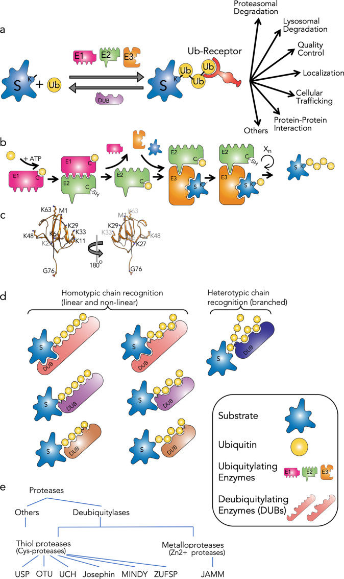
a A schematic representation of ubiquitylation/deubiquitylation events of target proteins and the cellular outcome. b The ubiquitylation cascade by three types of enzyme: ubiquitin-activating enzyme (E1), ubiquitin-conjugating enzyme (E2), and ubiquitin ligase (E3). c The seven lysine residues and the N-terminal methionine (M1) are projected on the structure of ubiquitin (PDB code 1UBQ). d Principles of substrate recognition and ubiquitin cleavage by DUBs. e. Families of DUBs classified by sequence homology of their catalytic domains.
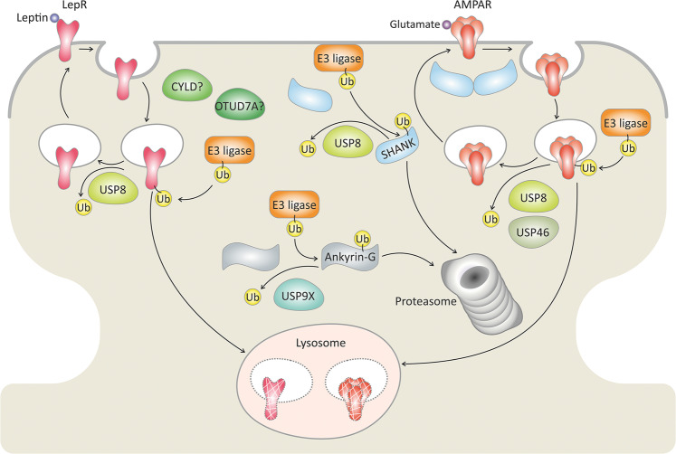
Activation of postsynaptic glutamate receptors controls neuron excitability. The postsynaptic density is enriched with a growing number of DUBs that antagonize E3-ligases and remove ubiquitin (Ub) chains, thereby protecting designated substrates from degradation by the lysosomes or the proteasome. Certain DUBs regulate the degradation and surface localization of the glutamate receptor, AMPAR. Moreover, a number of DUBs support the formation and function of the glutamatergic synapse by regulating the levels of scaffold proteins (SHANK and Ankyrin) and the leptin receptor (LepR). DUBs with yet unknown substrates are labeled with a question mark (?). The Ub symbol in this image indicates a polyubiquitin chain.
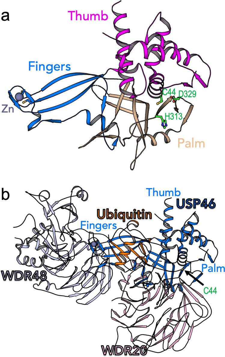
a The structure of USP46 subdomains. Residues of the catalytic triad located at the interface between the palm and the thumb subdomains are shown as green sticks (PDB code 5L8H). b A model of USP46 complex based on superposition of the structures of USP46 with covalent ubiquitin (Ub-VME) (PDB code 5L8H) with the non-covalent complex of USP46:WDR20:WDR48 (PDB code 6JLQ).
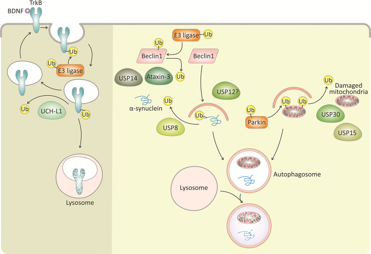
The cell survival tyrosine receptor kinase B (TrkB) is activated by BDNF and is ubiquitylated by E3-ligases for degradation. The DUB, UCH-L1, deubiqutylates TrkB and thereby regulates TrkB levels and surface localization. The accumulation of aggregate-prone proteins (for example, α-synuclein) and damaged mitochondria are associated with neuronal loss in neurodegenerative diseases, such as Parkinson’s disease. These proteins and damaged mitochondria are ubiquitylated by E3-ligases (for example, parkin ubiquitylation of damaged mitochondria) and are engulfed by autophagosomes for degradation in the lysosome. A number of DUBs antagonize autophagy-mediated degradation of damaged mitochondria (USP30 and USP15) and α-synuclein (USP8). Moreover, other DUBs (ataxin-3, USP14 and USP12) are involved in promoting neuronal autophagy. DUBs with yet unknown substrates are labeled with a question mark (?). The Ub symbol in this image indicates a polyubiquitin chain.
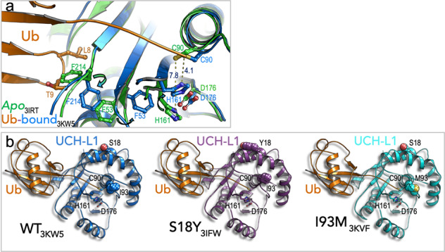
a Superposition of the structures of wild-type apo and covalent bound ubiquitin to UCH-L1 is shown (PDB code 3KW5). Active site rearrangement and activation is due to ubiquitin binding. b Structures of the Parkinson’s disease variants, S18Y and I93M, are shown (PDB codes 3IFW and 3KVF) in comparison to the structure of wild-type UCH-L1 in complex with covalent ubiquitin.
Similar articles
-
The pivotal role of ubiquitin-activating enzyme E1 (UBA1) in neuronal health and neurodegeneration.Int J Biochem Cell Biol. 2020 Jun;123:105746. doi: 10.1016/j.biocel.2020.105746. Epub 2020 Apr 18. Int J Biochem Cell Biol. 2020. PMID: 32315770 Review.
-
Distinct regulatory ribosomal ubiquitylation events are reversible and hierarchically organized.Elife. 2020 Feb 3;9:e54023. doi: 10.7554/eLife.54023. Elife. 2020. PMID: 32011234 Free PMC article.
-
A portrayal of E3 ubiquitin ligases and deubiquitylases in cancer.Int J Cancer. 2013 Dec 15;133(12):2759-68. doi: 10.1002/ijc.28129. Epub 2013 Mar 25. Int J Cancer. 2013. PMID: 23436247 Review.
-
Progressing neurobiological strategies against proteostasis failure: Challenges in neurodegeneration.Prog Neurobiol. 2017 Dec;159:1-38. doi: 10.1016/j.pneurobio.2017.08.005. Epub 2017 Sep 1. Prog Neurobiol. 2017. PMID: 28870769 Review.
-
La FAM fatale: USP9X in development and disease.Cell Mol Life Sci. 2015 Jun;72(11):2075-89. doi: 10.1007/s00018-015-1851-0. Epub 2015 Feb 12. Cell Mol Life Sci. 2015. PMID: 25672900 Free PMC article. Review.
Cited by
-
Enzymatically catalyzed molecular aggregation.Nat Commun. 2024 Nov 19;15(1):9999. doi: 10.1038/s41467-024-54291-1. Nat Commun. 2024. PMID: 39557870 Free PMC article.
-
Co-expression-wide association studies link genetically regulated interactions with complex traits.medRxiv [Preprint]. 2025 Jun 23:2024.10.02.24314813. doi: 10.1101/2024.10.02.24314813. medRxiv. 2025. PMID: 39711708 Free PMC article. Preprint.
-
Deubiquitinases as novel therapeutic targets for diseases.MedComm (2020). 2024 Dec 13;5(12):e70036. doi: 10.1002/mco2.70036. eCollection 2024 Dec. MedComm (2020). 2024. PMID: 39678489 Free PMC article. Review.
-
Ubiquitin Carboxyl-Terminal Hydrolase L1 and Its Role in Parkinson's Disease.Int J Mol Sci. 2024 Jan 21;25(2):1303. doi: 10.3390/ijms25021303. Int J Mol Sci. 2024. PMID: 38279302 Free PMC article. Review.
-
On the Study of Deubiquitinases: Using the Right Tools for the Job.Biomolecules. 2022 May 14;12(5):703. doi: 10.3390/biom12050703. Biomolecules. 2022. PMID: 35625630 Free PMC article. Review.
References
Publication types
MeSH terms
LinkOut - more resources
Full Text Sources
Other Literature Sources
Medical
