. 2021 Jan 22;7(1):5.
doi: 10.1038/s41540-020-00159-1.

A dynamic multi-tissue model to study human metabolism

Affiliations

A dynamic multi-tissue model to study human metabolism

Patricia Martins Conde et al. NPJ Syst Biol Appl. .

Abstract

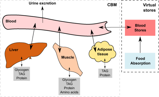
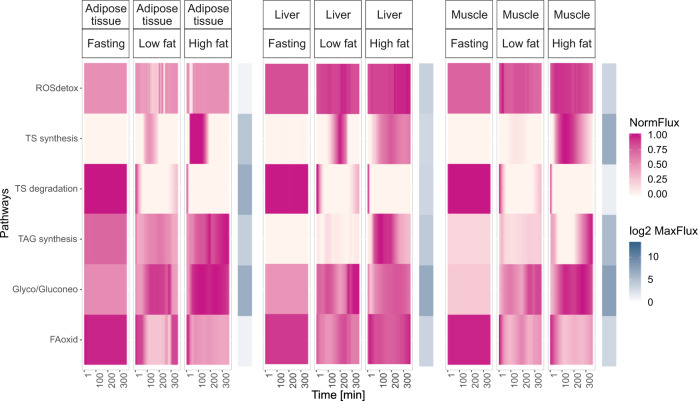
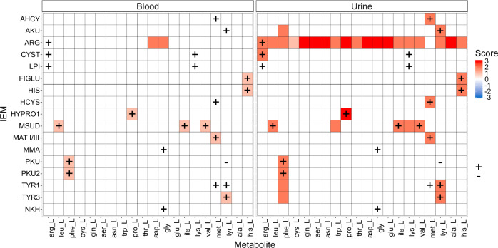
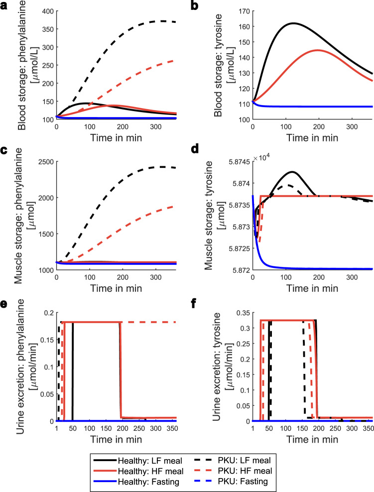
Metabolic modeling enables the study of human metabolism in healthy and in diseased conditions, e.g., the prediction of new drug targets and biomarkers for metabolic diseases. To accurately describe blood and urine metabolite dynamics, the integration of multiple metabolically active tissues is necessary. We developed a dynamic multi-tissue model, which recapitulates key properties of human metabolism at the molecular and physiological level based on the integration of transcriptomics data. It enables the simulation of the dynamics of intra-cellular and extra-cellular metabolites at the genome scale. The predictive capacity of the model is shown through the accurate simulation of different healthy conditions (i.e., during fasting, while consuming meals or during exercise), and the prediction of biomarkers for a set of Inborn Errors of Metabolism with a precision of 83%. This novel approach is useful to prioritize new biomarkers for many metabolic diseases, as well as for the integration of various types of personal omics data, towards the personalized analysis of blood and urine metabolites.


Conflict of interest statement

The authors declare no competing interests.

Figures

Fig. 1
Fig. 1. Model overview.
The multi-tissue model is divided in two parts. The first part, denoted here as CBM, contains the three tissue models, the blood compartment, and the urine excretion reactions. The second part, denoted here as virtual stores, contains the blood stores which are updated at each time step. The internal stores of each tissue are represented in grey with the stored compounds named in the boxes. The food absorption mimics the food absorbed by the gut and transferred to the blood. TAG = Triacylglycerol.
Fig. 2
Fig. 2. Metabolic pathways activity in different tissues associated with energy metabolism during fasting.
The pathway tissue storage degradation contains different reactions depending on each tissue. In the adipose tissue, this pathway contains the reaction that leads to the degradation of TAG from the adipose tissue stores. In the muscle, and in the liver, this pathway corresponds to the degradation of glycogen from the glycogen stores, and to the degradation of TAG from the fat stores in each tissue. The black arrow, occurring at around 2880 min, represents the point when the liver glycogen stores were depleted, leading to a switch in the metabolic activity. Each flux in a pathway was normalized to the maximum flux of that pathway for all tissues.
Fig. 3
Fig. 3. Comparison of the effect of different conditions on the energy associated pathways in different tissues.
The tissue storage degradation contains different reactions depending on the tissue. In the adipose tissue, this pathway contains the reaction that leads to the degradation of TAG from adipose tissue stores. In the muscle, and in the liver, this pathway corresponds to the degradation of glycogen from the glycogen stores, and to the degradation of TAG from the fat stores in each tissue. The tissue storage synthesis contains different reactions depending on each tissue. In the adipose tissue, this pathway contains the reaction that leads to the TAG synthesis, and its storage in the adipose tissue. In the muscle, and in the liver, this pathway corresponds to the synthesis, and storage of glycogen, and TAG in each tissue. Each flux in a pathway was normalized to the maximum flux of that pathway for each tissue. The fluxes were normalized in this way, as no striking pathway flux dynamics could be observed in the adipose tissue when the normalization was performed to the maximum value of a pathway among all tissues. ROSdetox = Reactive oxygen species (ROS) detoxification; TS synthesis = Tissue storage synthesis; TS degradation = Tissue storage degradation; TAG Synt = Triacylglycerol synthesis; Glyco/Gluconeo = Glycolysis, and gluconeogenesis; FAoxid = Fatty acid oxidation.
Fig. 4
Fig. 4. Prediction of amino acids biomarkers in different biofluids for a set of IEMs.
In red: metabolites predicted to have increased levels in an IEM. In blue: metabolites predicted to have decreased levels in an IEM. In white: metabolites predicted to remain unchanged in the presence of an IEM. Plus sign (+): metabolite level known to be increased in an IEM. Minus sign (−): metabolite level known to be decreased in an IEM. The data was taken from Shlomi et al. The score value represents the number of conditions, that the metabolite was predicted to be a biomarker in. As the IEMs were simulated in three different conditions, the absolute score value ranges from 0 to 3. With 0 representing a metabolite not identified as a biomarker, and a score of i.e., 3 representing a metabolite identified as being a biomarker in all 3 simulated conditions (fasting, low fat meal and high fat meal). Abbreviations of IEM names: AHCY = S-adenosylhomocysteine hydrolase; AKU = Alkaptonuria; ARG = Arginase deficiency; CYST = Cystinuria; LPI = Lysinuric Protein Intolerance; FIGLU = Glutamate Formiminotransferase Deficiency; HIS = Histidinemia; HCYS = Homocystinuria; HYPRO1 = Hyperprolinemia Type I; MSUD = Maple Syrup Urine Disease; MAT I/III = Methionine adenosyltransferase I/III deficiency; MMA= Methylmalonic Acidemia (MMA); PKU = Phenylketonuria; PKU2 = Phenylketonuria Type II; TYR1 = Tyrosinemia Type I; TYR3 = Tyrosinemia Type III; NKH = Glycine Encephalopathy/Nonketotic Hyperglycinemia.
Fig. 5
Fig. 5. Dynamic profile of phenylalanine and tyrosine during Phenylketonuria and healthy conditions.
LF meal = Low fat meal; HF meal = High fat meal; PKU = Phenylketonuria.

References

    1. Ma H, et al. The edinburgh human metabolic network reconstruction and its functional analysis. Mol. Syst. Biol. 2007;3:135. doi: 10.1038/msb4100177. - DOI - PMC - PubMed
    1. Duarte NC, et al. Global reconstruction of the human metabolic network based on genomic and bibliomic data. Proc. Natl Acad. Sci. USA. 2007;104:1777–1782. doi: 10.1073/pnas.0610772104. - DOI - PMC - PubMed
    1. Thiele I, et al. A community-driven global reconstruction of human metabolism. Nat. Biotechnol. 2013;31:419–425. doi: 10.1038/nbt.2488. - DOI - PMC - PubMed
    1. Swainston N, et al. Recon 2.2: from reconstruction to model of human metabolism. Metabolomics. 2016;12:109. doi: 10.1007/s11306-016-1051-4. - DOI - PMC - PubMed
    1. Brunk, E. et al. Recon 3 {D}: A resource enabling a three-dimensional view of gene variation in human metabolism. Nat. Biotechnol. (2017). - PMC - PubMed

Publication types

LinkOut - more resources