. 2021 Jan 22;11(1):2118.
doi: 10.1038/s41598-021-81253-0.

Discovery of a new class of integrin antibodies for fibrosis

Affiliations

Discovery of a new class of integrin antibodies for fibrosis

Ji Zhang et al. Sci Rep. .

Abstract

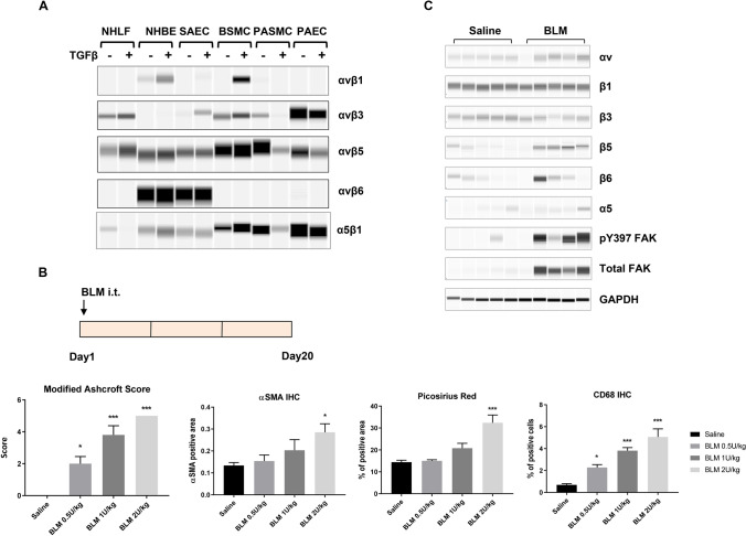
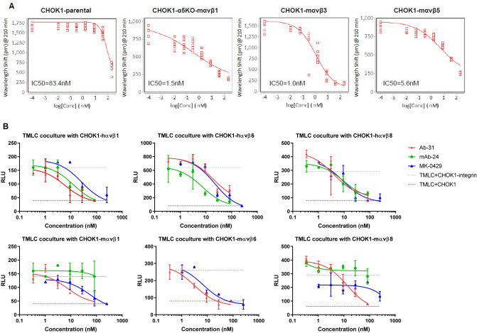
Lung fibrosis, or the scarring of the lung, is a devastating disease with huge unmet medical need. There are limited treatment options and its prognosis is worse than most types of cancer. We previously discovered that MK-0429 is an equipotent pan-inhibitor of αv integrins that reduces proteinuria and kidney fibrosis in a preclinical model. In the present study, we further demonstrated that MK-0429 significantly inhibits fibrosis progression in a bleomycin-induced lung injury model. In search of newer integrin inhibitors for fibrosis, we characterized monoclonal antibodies discovered using Adimab's yeast display platform. We identified several potent neutralizing integrin antibodies with unique human and mouse cross-reactivity. Among these, Ab-31 blocked the binding of multiple αv integrins to their ligands with IC50s comparable to those of MK-0429. Furthermore, both MK-0429 and Ab-31 suppressed integrin-mediated cell adhesion and latent TGFβ activation. In IPF patient lung fibroblasts, TGFβ treatment induced profound αSMA expression in phenotypic imaging assays and Ab-31 demonstrated potent in vitro activity at inhibiting αSMA expression, suggesting that the integrin antibody is able to modulate TGFβ action though mechanisms beyond the inhibition of latent TGFβ activation. Together, our results highlight the potential to develop newer integrin therapeutics for the treatment of fibrotic lung diseases.


Conflict of interest statement

The authors (J.Z., T.W., A.S., J.J., J.M., S.T., S.A.H., M.J.E., W.M., K.O., M.G-C., E.C-J., D.G.L, T.H., Q.Z., W.D., H.Y.M., J.H., T-Q.C., T.A., S.P., A.C.C., T.G., J.C.M., Z.R., S.T., H.H.S., M.H.) are/were employees of Merck Sharp & Dohme Corp., a subsidiary of Merck & Co., Inc., Kenilworth, NJ, USA and/or shareholders of Merck & Co., Inc., Kenilworth, NJ, USA.

Figures

Figure 1
Figure 1
Changes of αv integrin expression upon fibrosis induction in the lung. (A) The expression of αv integrins in various human primary lung cell types upon TGFβ (5 ng/ml for 24 h) treatment. Following immunoprecipitation with an anti-αv antibody, the αvβ1, αvβ3, αvβ5, and αvβ6 heterodimers were detected by Sally Sue simple western analysis after using antibodies that recognize each individual β-subunit. Normal human lung fibroblast, NHLF; normal human bronchial epithelial cells, NHBE; small airway epithelial cells, SAEC; bronchial smooth muscle cells, BSMC; pulmonary artery smooth muscle cells, PASMC; pulmonary artery endothelial cells, PAEC. Full-length blot images are presented in Supplemental Fig. S6A–E. (B) Development of a bleomycin-induced lung fibrosis model in mice. Bleomycin (BLM) was administered at the indicated doses via intra-tracheal (i.t.) instillation. After 20 days, lungs were collected for histological analyses. Modified Ashcroft score, Picosirus red staining, immunohistochemical analyses of αSMA and CD68 of total lung were quantified and shown (mean ± SEM, n = 5). Immunohistochemistry, IHC. One-way ANOVA followed by Tukey’s test, *p < 0.05, **p < 0.01, ***p < 0.005 vs Saline group. (C) Integrin expression and signaling in fibrotic lungs (BLM 0.5U/kg bw) was determined by Sally Sue simple western analysis using antibodies that recognized the individual α or β-subunits. Each lane represents total lung homegenate for one animal, n = 5 for saline or BLM group. GAPDH level in total lung lysates was used as a loading control. Full-length blot images are presented in Supplemental Fig. S6F–N.
Figure 2
Figure 2
MK-0429 inhibits lung fibrosis in the bleomycin mouse model. (A) Schematics of compound administration in BLM model. 5 days after BLM intra-tracheal instillation, the animals were given MK-0429 (200 mpk via osmotic minipump for 2 weeks) or Nintedanib (60 mpk po qd for 2 weeks). Lungs were collected at Day 19 for histological and biochemical evaluation. mpk, milligrams per kilogram body weight; po, Per os, oral administration; qd, Quaque die, every day. (B) Plasma total drug concentration was measured 2 h after final oral dose at Day 19. (C) Representative Masson Trichrome staining of mouse lungs. a and d, saline intra-tracheal instillation; b-f, BLM intra-tracheal instillation; a and d, no compound treatment; b, vehicle in minipump; c, MK-0429 in minipump; e, vehicle po; f, Nintedanib po. D) Modified Ashcroft scores of mouse lung. Mean ± SEM, n = 10. One-way ANOVA followed by Tukey’s test, *p < 0.05, **p < 0.01, ***p < 0.005 vs BLM-vehicle group. (E) Total inflammation area in mouse lungs. Mean ± SEM, n = 10. One-way ANOVA followed by Tukey’s test, *p < 0.05, **p < 0.01, ***p < 0.005 vs BLM-vehicle group. (F) Immunohistochemical analysis of αSMA. Mean ± SEM, n = 10. One-way ANOVA followed by Tukey’s test, *p < 0.05, **p < 0.01, ***p < 0.005 vs BLM-vehicle group. (G) Soluble collagen content in bronchoalveolar lavage fluid (BALF). Mean ± SEM, n = 10. *p < 0.05, **p < 0.01, ***p < 0.005 vs BLM-vehicle group.
Figure 3
Figure 3
Integrin antibody screening and assay development. (A) Staged efforts to screen integrin antibodies from human naïve IgG library by using Adimab’s yeast display platform. (B) IgG-expressing yeast clone selection process. The X-axis represents integrin binding and the Y-axis reflects antibody expression. Yeast cell population with strong antigen binding (boxed) was sorted out for the next round of selection. (C) Cell-based ELISA (CELISA) binding assays were used as the primary screen for integrin antibody selection. Dose-dependent binding of the benchmarking integrin antibody mAb-24 to various integrin-expressing CHOK1 cells was shown. (D) AlphaLISA integrin-ligand binding assays were used for in vitro functional screen. Dose-dependent inhibition of human integrin-ligand binding by MK-0429 in AlphaLISA assay panel.
Figure 4
Figure 4
Discovery a set of antibodies with strong blocking activities against both human and mouse αv integrins. (A) Titration of Ab-29, Ab-30, Ab-31, Ab-32, and Ab-33 for their binding to CHOK1-human αvβ1, αvβ6, and α5β1 stable cell lines in CELISA assays. (B) EC50 of Ab-29, Ab-30, Ab-31, Ab-32, and Ab-33 in CELISA assays, as well as their IC50 in human and mouse AlphaLISA integrin blocking assays.
Figure 5
Figure 5
Ab-31 inhibits integrin-mediated cell adhesion and latent TGFβ activation. (A) The effect of Ab-31 on the adhesion of CHOK1 parental, CHOK1-α5KO-mαvβ1, CHOK1-mαvβ3, and CHOK1-mαvβ5 cells to fibronectin or vitronectin matrix. (B) pan-αv integrin inhibitors suppress latent TGFβ activation in the transfected Mink lung epithelial cells (TMLC) and CHOK1-integrin co-culture system. The effects of Ab-31, MK-0429, and benchmarking mAb-24 on PAI-1 luciferase activity were shown.
Figure 6
Figure 6
Ab-31 reduces TGFβ-induced αSMA expression in lung fibroblasts. (A) Normal human lung fibroblasts were stimulated with TGFβ (5 ng/mL) and stained with anti-αSMA antibody for immunofluorescence analysis. Cells were pre-treated with or without MK-0429 (10 μM) or the ALK5 inhibitor SB-525334 (10 μM) 30 min before the addition of TGFβ. 48 h after the treatment, cells were imaged with Opera Phenix high-content screening system for αSMA expression (fluorescence intensity) and αSMA-associated morphological changes (STAR program). (B) The effects of Ab-31, MK-0429, and benchmarking antibody mAb-24 on TGFβ-associated αSMA induction in IPF patient lung fibroblasts. C) Structural modeling predicts a distinct integrin binding mode for Ab-31. The structures of two therapeutic monoclonal antibodies, Abituzumab (17E6, RCSB PDB: 4O02) and LM609 (RCSB PDB: 6AVQ), in complex with αvβ3 integrin were shown to highlight the difference in binding modes for each molecule. A model of Ab-31 and αvβ3 integrin complex was determined by docking of related antibody sequence to αvβ3 structure (see “Methods”). For visualization, only the Fv region of each antibody were shown.

Similar articles

Cited by

References

    1. Wilson KC, Raghu G. The 2015 guidelines for idiopathic pulmonary fibrosis: An important chapter in the evolution of the management of patients with IPF. Eur. Respir. J. 2015;46:883–886. doi: 10.1183/13993003.01335-2015. - DOI - PubMed
    1. Hynes RO. Integrins: Bidirectional, allosteric signaling machines. Cell. 2002;110:673–687. doi: 10.1016/s0092-8674(02)00971-6. - DOI - PubMed
    1. Munger JS, et al. The integrin alpha v beta 6 binds and activates latent TGF beta 1: A mechanism for regulating pulmonary inflammation and fibrosis. Cell. 1999;96:319–328. doi: 10.1016/s0092-8674(00)80545-0. - DOI - PubMed
    1. Kitamura H, et al. Mouse and human lung fibroblasts regulate dendritic cell trafficking, airway inflammation, and fibrosis through integrin alphavbeta8-mediated activation of TGF-beta. J. Clin. Invest. 2011;121:2863–2875. doi: 10.1172/JCI45589. - DOI - PMC - PubMed
    1. Reed NI, et al. The alphavbeta1 integrin plays a critical in vivo role in tissue fibrosis. Sci. Transl. Med. 2015;7:288. doi: 10.1126/scitranslmed.aaa5094. - DOI - PMC - PubMed

Publication types

MeSH terms

Substances