. 2021 Jan 22;11(1):2097.
doi: 10.1038/s41598-021-81639-0.

Loss of Parkinson's susceptibility gene LRRK2 promotes carcinogen-induced lung tumorigenesis

Affiliations

Loss of Parkinson's susceptibility gene LRRK2 promotes carcinogen-induced lung tumorigenesis

Chandra Lebovitz et al. Sci Rep. .

Abstract

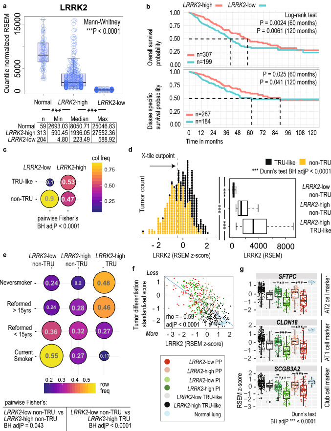
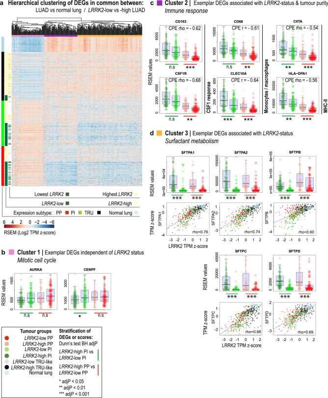
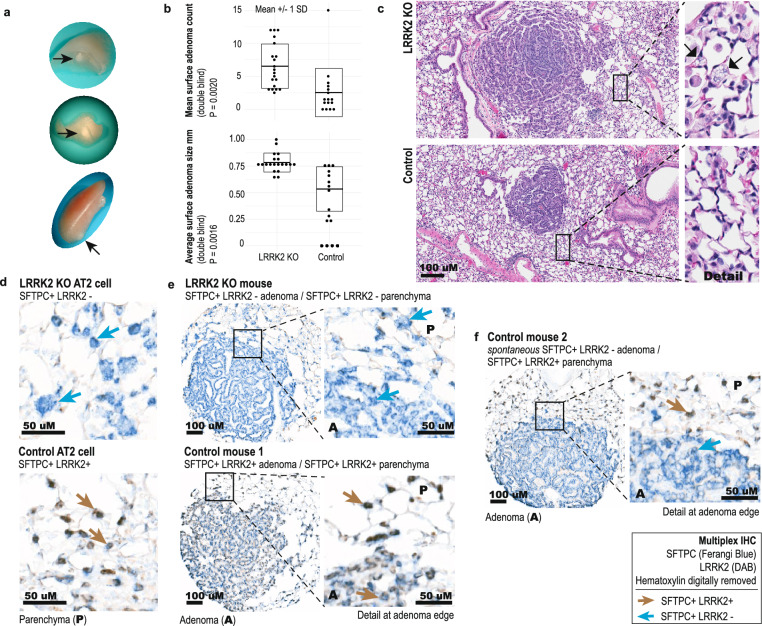
Pathological links between neurodegenerative disease and cancer are emerging. LRRK2 overactivity contributes to Parkinson's disease, whereas our previous analyses of public cancer patient data revealed that decreased LRRK2 expression is associated with lung adenocarcinoma (LUAD). The clinical and functional relevance of LRRK2 repression in LUAD is unknown. Here, we investigated associations between LRRK2 expression and clinicopathological variables in LUAD patient data and asked whether LRRK2 knockout promotes murine lung tumorigenesis. In patients, reduced LRRK2 was significantly associated with ongoing smoking and worse survival, as well as signatures of less differentiated LUAD, altered surfactant metabolism and immunosuppression. We identified shared transcriptional signals between LRRK2-low LUAD and postnatal alveolarization in mice, suggesting aberrant activation of a developmental program of alveolar growth and differentiation in these tumors. In a carcinogen-induced murine lung cancer model, multiplex IHC confirmed that LRRK2 was expressed in alveolar type II (AT2) cells, a main LUAD cell-of-origin, while its loss perturbed AT2 cell morphology. LRRK2 knockout in this model significantly increased tumor initiation and size, demonstrating that loss of LRRK2, a key Parkinson's gene, promotes lung tumorigenesis.


Conflict of interest statement

The authors declare no competing interests.

Figures

Figure 1
Figure 1
LRRK2-low lung adenocarcinoma is associated with poor patient survival, non-TRU expression-based molecular subtypes and worse predicted tumor differentiation. (A) Plot of LRRK2 mRNA levels in LUAD tumors (dichotomized into LRRK2-low and -high expression groups) and adjacent normal lung tissue (RSEM values), from TCGA LUAD patients. (B) Kaplan–Meier plot of LUAD patient OS or DSS stratified by LRRK2 expression status. (C) Pairwise Fisher’s exact test for enrichment of expression subtype frequency within LRRK2 expression groups: TRU-like versus non-TRU type LUAD. (D) (Left) Standardized LRRK2 tumoral expression per sample (RSEM values), annotated for lower risk TRU-like or higher risk non-TRU type tumors or (Right) grouped by the combined status for LRRK2 expression and expression subtype (median centred boxplot of RSEM values). (E) Pairwise Fisher’s exact test for enrichment of smoking history within LRRK2 expression groups: TRU-like versus non-TRU type LUAD. (F) Correlation of LRRK2 expression with a previously published gene expression-based score representing LUAD tumor differentiation status (Spearman’s correlation coefficient − 0.59 with Holm’s adjP < 0.0001; positive scores represent increasingly poor differentiation). (G) Stratification of the tumoral gene expression of established markers for alveolar and bronchiolar epithelial cell types, by the combined LRRK2 and expression subtype status of LUAD tumors (median-centred boxplot of standardized RSEM values; Dunn’s test BH adjP < 0.05).
Figure 2
Figure 2
The transcriptional landscape of LRRK2 repression in lung adenocarcinoma patients. (A) Heatmap depicting the hierarchical agglomerative clustering of genes differentially expressed in common between two biological contexts in TCGA LUAD tumors, each with a marked reduction of LRRK2 expression: (1) across all tumors versus normal lung and (2) in the lowest LRRK2 expressing tumors versus the highest LRRK2 expressing tumors. Columns: n = 385 DEGs with absolute fold change of median RSEM value > 2; Rows: n = 59 normal lung and n = 517 LUAD tumors, ordered by tissue, LRRK2 expression status and expression subtype. (B) Exemplar DEGs identified in Cluster 1, enriched for genes that act in mitotic cell cycle (Metascape algorithm; q < 0.05), stratified by tumor group (multicolour boxplots of exemplar gene expression; Dunn’s test BH adjP > 0.05). Tumor groups represent the combined sample status for LRRK2 expression and non-TRU subtype. (C) Exemplar DEGs identified in Cluster 2, associated with tumor purity (consensus purity estimate or CPE; Spearman’s correlation coefficient ≥ 0.5 with Holm’s corrected P < 0.0001) and enriched for immune response genes (Metascape algorithm; q < 0.05), stratified by tumor group (multicolour boxplots of exemplar gene expression; Dunn’s test BH adjP < 0.01). (D) Exemplar DEGs identified in Cluster 3, enriched for genes that act in surfactant metabolism (Metascape algorithm; q < 0.05), stratified by tumor group (multicolour boxplots of exemplar gene expression; Dunn’s test BH adjP < 0.001) or plotted against LRRK2 expression (multicolour scatterplots; Spearman’s correlation coefficient ≥ 0.6 with Holm’s corrected P < 0.0001; standardized RSEM Transcripts Per Million or TPM).
Figure 3
Figure 3
LRRK2 knockout increases SFTPC-positive tumor initiation in a murine model of urethane-induced, early stage lung cancer. (A) Representative visible adenomas that developed in the lungs of both LRRK2 knockout (KO) mice and wild type (WT) strain mates, following urethane treatment. (B) Double blind analysis of adenoma counts and size (n = 16 WT and n = 20 LRRK2 KO animals, with n = 40 WT and n = 130 LRRK2 KO surface lesions). (C) Representative H&E stained sections of adenomas (20× magnification; transverse section; n = 16 WT and n = 20 LRRK2 KO animals, with n = 40 WT and n = 130 LRRK2 KO surface lesions). (Inset) Detailed view of representative vacuolated cells (arrows) found in LRRK2 KO lung only. (D) Representative images of multiplexed IHC staining for colocalization of alveolar type II (AT2)-specific cell marker SFTPC (Ferangi blue chromogen) and LRRK2 (3,3′-Diaminobenzidine (DAB) chromogen), in LRRK2 KO (top) and WT (bottom) mouse lung parenchyma (n = 7 WT mice with 14 lesions; n = 15 KO mice with 25 lesions). (E) Representative images of multiplexed IHC staining for SFTPC and LRRK2, in adenomas that developed in the lungs of both LRRK2 KO (top) and WT (bottom) control mice, following urethane treatment (n = 7 WT mice with 14 lesions; n = 15 KO mice with 25 lesions). (Inset) detailed view of staining at adenoma edge and adjacent lung parenchyma. (F) Representative images of multiplexed IHC staining for SFTPC and LRRK2, for adenomas that developed in the lungs of WT control mice following urethane treatment, showing spontaneous reduction of LRRK2 positivity. Representative of 4 of 14 lesions from n = 7 WT control mice. (Inset) AT2 cells in the adjacent lung parenchyma remained double positive for SFTPC and LRRK2.

References

    1. World Health Organization. Cancer key facts. https://www.who.int/news-room/fact-sheets/detail/cancer (2018).
    1. Alderton GK. Tumour evolution: Clonal ancestry in lung cancer. Nat. Rev. Cancer. 2014;14:763. doi: 10.1038/nrc3867. - DOI - PubMed
    1. Lebovitz CB, et al. Cross-cancer profiling of molecular alterations within the human autophagy interaction network. Autophagy. 2015;11:1668–1687. doi: 10.1080/15548627.2015.1067362. - DOI - PMC - PubMed
    1. Feng DD, Cai W, Chen X. The associations between Parkinson’s disease and cancer: The plot thickens. Transl. Neurodegener. 2015;4:20. doi: 10.1186/s40035-015-0043-z. - DOI - PMC - PubMed
    1. Kluss JH, Mamais A, Cookson MR. LRRK2 links genetic and sporadic Parkinson’s disease. Biochem. Soc. Trans. 2019 doi: 10.1042/BST20180462. - DOI - PMC - PubMed

Publication types

MeSH terms

Substances