. 2021 Jan 22;4(1):104.
doi: 10.1038/s42003-020-01623-8.

Chromosomal scale assembly of parasitic wasp genome reveals symbiotic virus colonization

Affiliations

Chromosomal scale assembly of parasitic wasp genome reveals symbiotic virus colonization

Jérémy Gauthier et al. Commun Biol. .

Erratum in

  • Author Correction: Chromosomal scale assembly of parasitic wasp genome reveals symbiotic virus colonization.
    Gauthier J, Boulain H, van Vugt JJFA, Baudry L, Persyn E, Aury JM, Noel B, Bretaudeau A, Legeai F, Warris S, Chebbi MA, Dubreuil G, Duvic B, Kremer N, Gayral P, Musset K, Josse T, Bigot D, Bressac C, Moreau S, Periquet G, Harry M, Montagné N, Boulogne I, Sabeti-Azad M, Maïbèche M, Chertemps T, Hilliou F, Siaussat D, Amselem J, Luyten I, Capdevielle-Dulac C, Labadie K, Merlin BL, Barbe V, de Boer JG, Marbouty M, Cônsoli FL, Dupas S, Hua-Van A, Le Goff G, Bézier A, Jacquin-Joly E, Whitfield JB, Vet LEM, Smid HM, Kaiser L, Koszul R, Huguet E, Herniou EA, Drezen JM. Gauthier J, et al. Commun Biol. 2021 Jul 30;4(1):940. doi: 10.1038/s42003-021-02480-9. Commun Biol. 2021. PMID: 34331006 Free PMC article. No abstract available.

Abstract

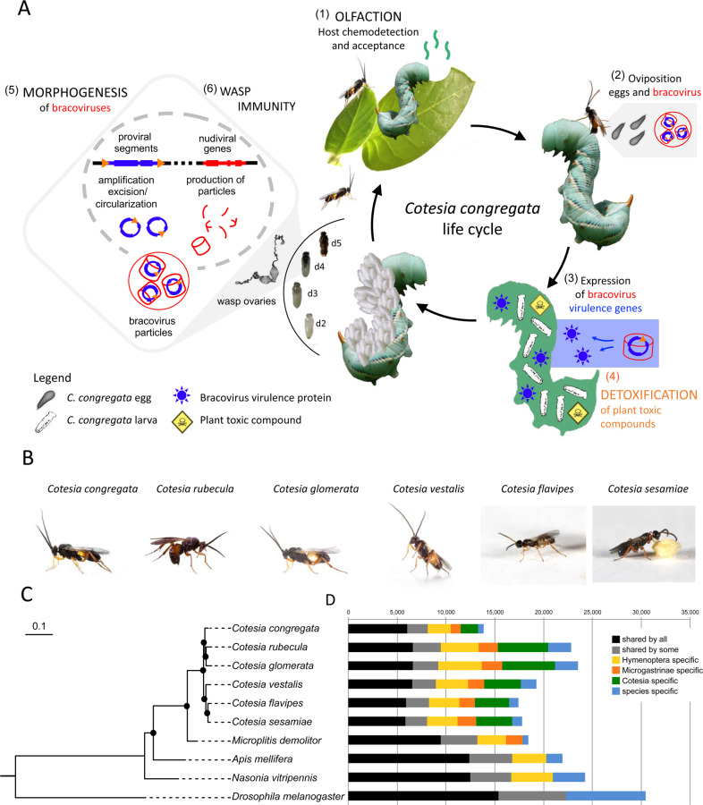
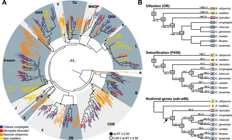
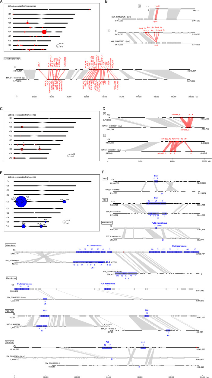
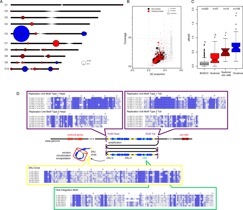
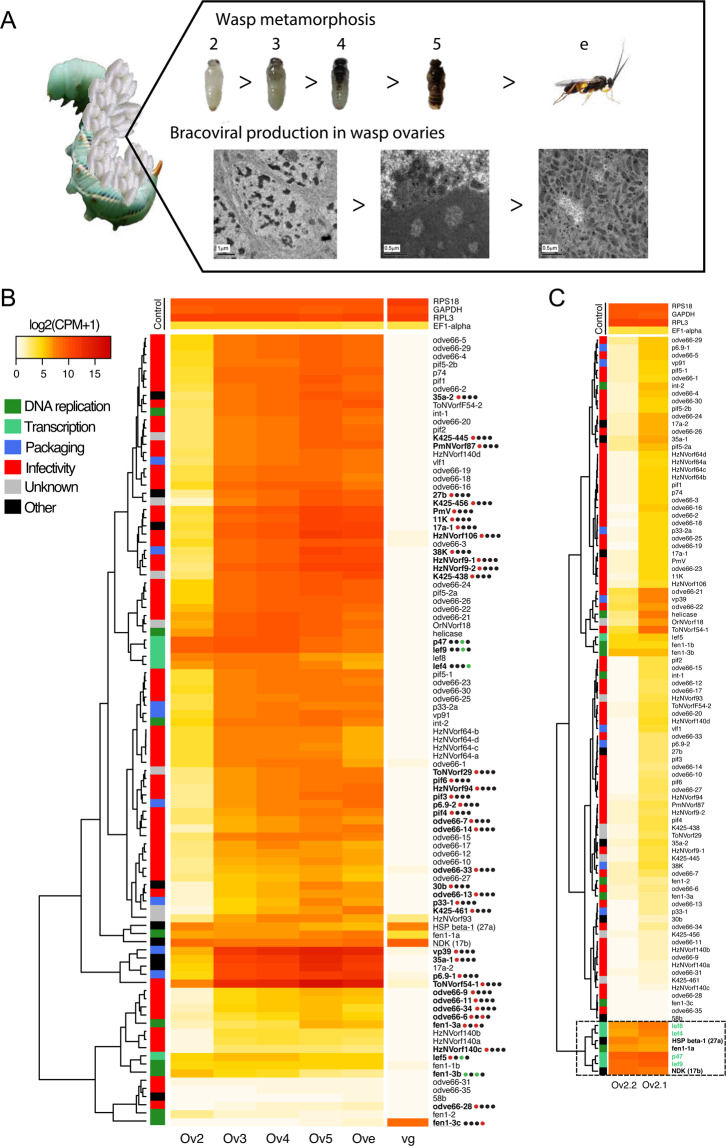
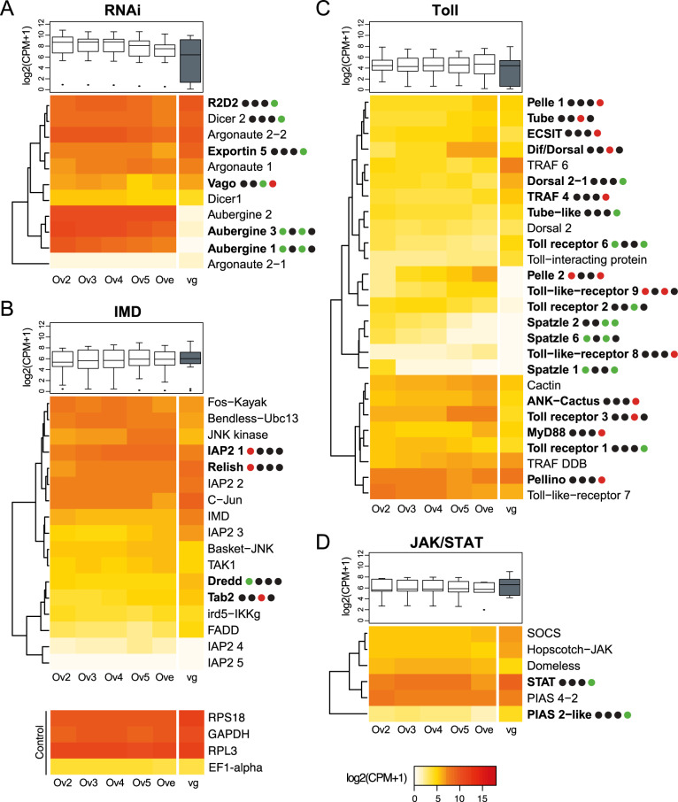
Endogenous viruses form an important proportion of eukaryote genomes and a source of novel functions. How large DNA viruses integrated into a genome evolve when they confer a benefit to their host, however, remains unknown. Bracoviruses are essential for the parasitism success of parasitoid wasps, into whose genomes they integrated ~103 million years ago. Here we show, from the assembly of a parasitoid wasp genome at a chromosomal scale, that bracovirus genes colonized all ten chromosomes of Cotesia congregata. Most form clusters of genes involved in particle production or parasitism success. Genomic comparison with another wasp, Microplitis demolitor, revealed that these clusters were already established ~53 mya and thus belong to remarkably stable genomic structures, the architectures of which are evolutionary constrained. Transcriptomic analyses highlight temporal synchronization of viral gene expression without resulting in immune gene induction, suggesting that no conflicts remain between ancient symbiotic partners when benefits to them converge.


Conflict of interest statement

The authors declare no competing interests.

Figures

Fig. 1
Fig. 1. Cotesia species life cycle, phylogeny, and gene content.
A Major traits involved in the parasitoid koinobiont lifestyle and genome content of six Cotesia species. First (1) OLFACTION plays an important role in the detection of the plant (tobacco) attacked by caterpillars and host (M. sexta) larvae acceptance by adult wasps (C. congregata). Once the host is accepted, the wasp injects its eggs bathed in ovarian fluid filled with bracovirus particles (2). Bracovirus particles infect host cells, from which expression of bracovirus virulence genes (3) alter host immune defenses, allowing wasp larvae development (the eggs laid in the host body would otherwise be engulfed in a cellular sheath of hemocytes). As the host ingests plant toxic compounds, such as nicotine, while feeding, wasp larvae consuming the hemolymph containing these compounds rely on (4) detoxification to complete their life cycle. However, in these species associated with endogenous viruses the most important trait for parasitism success consists in (5) bracovirus morphogenesis during wasp metamorphosis, using genes originating from a nudivirus ancestrally integrated in the wasp genome. As massive production of virus particles occurs within wasp ovaries, (6) wasp immunity may be induced during particles production; d2, d3, d4, d5 refer to developmental stages of C. congregata larvae. B Pictures of the six Cotesia species sequenced (credit H. M. Smid and R. Copeland). C Phylogeny of these species based on 1058 single-copy orthologous insect genes including the Microgastrinae Microplitis demolitor and outgroups (N. vitripennis, A. mellifera, and D. melanogaster). Black dots highlight branches with at least 90% support from maximum-likelihood analysis (1000 bootstraps). D Distribution of shared genes at several phylogenetic levels. Full protein-coding gene sets were included to identify orthologous gene groups. The “shared by some” category refers to genes shared by at least nine species among the ten studied. Note that the lower number of genes for C. congregata probably reflects the higher quality of the genome assembly obtained.
Fig. 2
Fig. 2. Gene family extensions in Cotesia.
A Maximum-likelihood phylogeny of the OR family in C. congregata and four other Hymenoptera species. The dataset included 243 amino acid sequences from C. congregata (blue), 203 sequences from M. demolitor (red), 216 sequences from N. vitripennis (orange), 162 sequences from A. mellifera (yellow). The tree was rooted using the Orco (OR-coreceptor) clade. Circles indicate nodes strongly supported by the approximate likelihood-ratio test (black circles aLRT ≥ 0.95; white circles 0.90 ≥ aLRT ≤ 0.95). The scale bar represents 0.5 expected amino acid substitutions per site. ORs of the five Hymenoptera species are distributed into 18 OR subfamilies previously described in delineated in gray. B Copy number dynamics of OR (olfaction) P450 (detoxification) and Odv-e66 genes, note that the later are found specifically in bracovirus-associated wasps since they derive from the ancestrally integrated nudivirus. Estimated numbers of gene gain and loss events are shown on each branch of the species tree. The size of OR repertoires in common ancestors is indicated in the boxes. The lack of phylogenetic resolution for closely related Cotesia OR genes precluded any comprehensive analysis of gene gains and losses.
Fig. 3
Fig. 3. Synteny of nudiviral genes loci and proviral loci (PL) between C. congregata and M. demolitor.
A C. congregata chromosome map with the position of 24 nudiviral genes loci. B Comparisons between nudiviral gene regions of C. congregata and M. demolitor. Synteny between the two species has been characterized by at least two hymenopteran (non-viral) orthologous genes in the vicinity of homologous nudiviral gene(s) of both species. Genome scaffolds are represented in black. Red boxes indicate nudiviral genes and white boxes refer to hymenopteran genes. 1. the vp91 region is orthologous indicating the position of this gene was inherited from their common ancestor 53 mya; 2. the fen region is also orthologous but an expansion occurred specifically in Cotesia lineage giving rise to six copies; 3. the organization of the nudiviral cluster encoding in particular capsid genes has remained strikingly similar with the same viral genes in the same order (except p6-9-2) in both species indicating strong evolutionary constraints. C C. congregata chromosome map with the position of gene loci corresponding to the highly expanded odv-e66 nudiviral gene family. D Comparison of two odv-e66 loci showing that expansion occurred before (cluster 7) and after (cluster 4) the separation of both species E C. congregata chromosome map with the position of Proviral Loci (PL) encoding virulence genes packaged in bracovirus particles. Note the concentration of loci (successively PL10-PL1-PL2 and PL4) in a 2 Mb region termed “macrolocus” and representing half of the chromosome 5 short arm. F Comparison of C. congregata and M. demolitor PL. Numbers 1 to 37 and letters correspond to the different dsDNA circles present in CcBV and MdBV particles produced from the PL. Blue boxes indicate virulence genes while white boxes refer to hymenopteran genes and the red boxes to a nudiviral odv-e66 gene located between PL1 and PL2 and 58b near PL3-PL7. Ø indicates the absence of orthologs PL in the M. demolitor genome.
Fig. 4
Fig. 4. Bracovirus genes and motifs architecture and evolution.
A C. congregata chromosome map with the location of all bracovirus loci: nudiviral gene loci are shown in red, nudiviral odv-e66 gene loci in hatched red and Proviral Loci (PL) in blue. The sizes of the circles correspond to the relative number of genes in each locus B Taxon-annotated GC content-coverage plot of the C. congregata genome associated with Braconidae (in black) and Polydnaviridae (in red). Each circle represents a scaffold in the assembly, scaled by length, and colored by taxonomy assigned by BlobTools. The x-axis corresponds to the average GC content of each scaffold and the y-axis corresponds to the average coverage based on alignment of the Illumina reads. C Measure of selection pressure on hymenopteran conserved genes, nudiviral genes and virulence genes. Pairwise evolutionary rates (dN/dS) of single-copy orthologous BUSCO genes, nudiviral genes, different copies from the expanded odv-e66 nudiviral gene family and virulence genes of C. congregata and C. sesamiae. Letters above boxes indicate significant differences determined by Kruskal–Wallis test (H = 296.8, 2 d.f., P < 0.001) followed by post hoc comparisons. D Schematic representation of the genomic amplification during the production of viral particles in the wasp ovaries. Replication Unit Motifs (RUM) are the motifs that constitute the extremities of the molecules amplified during particle production. Direct repeat junctions (DRJ), at the extremities of each segment are used during the excision/circularization process to produce packaged dsDNA circles from the amplified molecules. Host integration motifs (HIM) are motifs used during the integration of bracovirus circles in host genome. For each of these motifs an alignment of a representative set of sequence comprising five motifs from C. congregata and M. demolitor are represented (complete alignments are shown in Supplementary Fig. 4).
Fig. 5
Fig. 5. Gene expression of nudiviral genes in the ovaries during C. congregata nymphal development.
A Pictures of wasp developmental stages studied and characteristic electron micrographs of ovarian cells involved in particle production. From d2 to d4 stage, cells that will produce particles show enlarged nuclei with chromatin condensation (left panel). From d5 massive particle production begins, particle assembly occurs at the periphery of a zone of electron dense material in the nucleus named “virogenic stroma” (middle panel). In newly emerged wasp nuclei are completely filled with bracovirus particles (right panel). Credit: Juline Herbinière. B Unsupervised hierarchical clustering based on gene expression in ovaries and venom glands. Ov2, Ov3, Ov4, Ov5 refer to the different ovaries across wasp developmental stages. Ove and vg refer to ovaries and venom glands of adult wasps. The colored squares associated with the clustering tree indicate the viral functions to which different nudiviral genes are supposed to contribute based on those of their baculovirus homologs. Heatmap of expression levels of 95 nudiviral genes is shown in the middle panel. Bold names highlight the genes that are validated as significantly differentially expressed between two consecutive stages using the statistical analysis and dots represent the four different comparisons studied between ovary stages (Ov2 vs. Ov3, Ov3 vs. Ov4, Ov4 vs. Ov5 and Ov5 vs. Ove). Black, red, and green dots indicate similar, increased and reduced expressions between consecutive developmental stages, respectively. The increase of some nudivirus genes expression between d2 and d3 visualized on the heat map was not validated statistically for all of them because in one of the d2 duplicates (shown in C) nudiviral genes expression had already reached high levels. Underlined genes show higher expression in venom glands compared to ovaries (Ove) note that 27a and 17b are not nudiviral genes but wasp genes, the products of which have been identified in Chelonus inanitus bracovirus particles. The expression of four stable wasp genes having high (RPS18, RPL3, GAPDH) or low expression level (EF1-alpha) is presented as control for comparison. C Unsupervised hierarchical clustering of gene expression from the two replicates of Ov2 ovary stage, that are very different regarding nudiviral gene expression levels, although dissected nymphae presented a similar coloration pattern, the left one representing a slightly earlier stage from the analysis of the whole set of wasp genes. Note that the genes within the box are already expressed at a high level in the earlier stage, including all of the genes involved in nudiviral transcription (shown in green) except lef5, in accordance with the hypothesis that the nudiviral RNA polymerase complex controls the expression of the other genes (lef-5 is associated with the complex but not a part of it).
Fig. 6
Fig. 6. Gene expression of antiviral immune genes in the ovaries during C. congregata nymphal development.
The heatmaps show the expression of genes involved in A RNAi, B Imd, C Toll and D Jak-STAT pathways across the developmental stages of ovaries (Ov2, Ov3, Ov4, Ov5, Ove) and in venom glands (vg). The trees on the left are unsupervised hierarchical clustering of expression values. Boxplots represent overall expression of each pathway in ovaries and venom glands. Bold names highlight the genes that are differentially expressed between two stages and dots represent the four different comparisons studied between consecutive ovary stages (Ov2 vs. Ov3, Ov3 vs. Ov4, Ov4 vs. Ov5 and Ov5 vs. Ove). Black, red, and green dots indicate similar, increased and reduced expressions between consecutive developmental stages, respectively. Note that no particular trend appears correlated to bracovirus particles production, which occurs massively from Ov5 onward. The expression of four stable wasp genes having high (RPS18, RPL3, GAPDH) or low expression level (EF1-alpha) is presented as control for comparison.

References

    1. Parra JRP. biological control in Brazil: an overview. Sci. Agric. 2014;71:420–429.
    1. Parra JRP, Coelho A. Applied biological control in Brazil: from laboratory assays to field application. J. Insect Sci. 2019;19:1–6. - PMC - PubMed
    1. Dicke M. Behavioural and community ecology of plants that cry for help. Plant Cell Env. 2009;32:654–666. - PubMed
    1. Poelman EH, et al. Hyperparasitoids use herbivore-induced plant volatiles to locate their parasitoid host. PLoS Biol. 2012;10:e1001435. - PMC - PubMed
    1. Gauthier J, Drezen JM, Herniou EA. The recurrent domestication of viruses: major evolutionary transitions in parasitic wasps. Parasitology. 2018;145:713–723. - PubMed

Publication types

Substances

LinkOut - more resources