. 2021 Jan;5(1):89-102.
doi: 10.1038/s41551-020-00674-w. Epub 2021 Jan 22.

Mechanobiological conditioning of mesenchymal stem cells for enhanced vascular regeneration

Affiliations

Mechanobiological conditioning of mesenchymal stem cells for enhanced vascular regeneration

Jason Lee et al. Nat Biomed Eng. 2021 Jan.

Abstract

Using endogenous mesenchymal stem cells for treating myocardial infarction and other cardiovascular conditions typically results in poor efficacy, in part owing to the heterogeneity of the harvested cells and of the patient responses. Here, by means of high-throughput screening of the combinatorial space of mechanical-strain level and of the presence of particular kinase inhibitors, we show that human mesenchymal stem cells can be mechanically and pharmacologically conditioned to enhance vascular regeneration in vivo. Mesenchymal stem cells conditioned to increase the activation of signalling pathways mediated by Smad2/3 (mothers against decapentaplegic homolog 2/3) and YAP (Yes-associated protein) expressed markers that are associated with pericytes and endothelial cells, displayed increased angiogenic activity in vitro, and enhanced the formation of vasculature in mice after subcutaneous implantation and after implantation in ischaemic hindlimbs. These effects were mediated by the crosstalk of endothelial-growth-factor receptors, transforming-growth-factor-beta receptor type 1 and vascular-endothelial-growth-factor receptor 2. Mechanical and pharmacological conditioning can significantly enhance the regenerative properties of mesenchymal stem cells.


Figures

Figure 1.
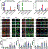
Figure 1.. Specific types of mechanical stretch activate Smad2/3 and Yap/Taz pathways in mesenchymal stem cells.
(A) Transcription factor activity in MSCs was measured using a luciferase reporter assay after the application of cyclic mechanical strain (5% maximal strain) for 8 hours with co-treatment with 10 ng/ml VEGF-A or 10 ng/ml TGF-β1 (n = 8). *p = 0.001 versus static control group. p = 0.001 versus static, growth factor treated group. p = 0.024 versus control group under the same mechanical loading conditions. (B) Smad transcription factor activity in MSCs with application of load for 24 hours using the multi-strain configuration (n = 6). *p = 0.024 versus static control group. p = 0.038 versus static, growth factor treated group. p = 0.049 versus control group under the same mechanical loading conditions. (C) The MSCs were treated with mechanical load using the multi-strain format at 0.1 Hz for 24 hours and then immunostained for markers of vascular cell differentiation or signaling pathway activation. Scale bar = 100 μm. (D) Ratio of nuclear to cytoplasmic p-Smad2/3 in MSC after mechanical loading for 24 hours. *p = 0.02 versus static group. (E) Ratio of nuclear to cytoplasmic Yap/Taz in the mechanically loaded MSCs (n = 4). *p = 0.039 versus static group. (F) Quantification of PECAM and α-SMA staining in MSCS after 24 hours of mechanical loading at 0.1 Hz (n = 4). *p = 0.005 versus static group. (G) Strain waveforms for sine and brachial loading at 0.1 Hz. (H) Quantification of western blotting for vascular markers and signal activation after 24 hours of mechanical loading (n = 4). *p = 0.01 versus static and sine groups.
Figure 2.
Figure 2.. High throughput mechanobiological screen for small molecule inhibitors that have synergistic activation of Yap/Taz and Smad2/3 with mechanical loading.
The MSCs were treated with 7.5% mechanical strain at 0.1 Hz for 24 hours in the presence of compounds from a library of kinase inhibitors. (A) The cells were immunostained and quantified for nuclear localization of Yap/Taz and p-Smad2/3 (n = 5). *p = 0.037 versus cells treated with static conditions under control treatment. p = 0.001 versus mechanically strained control group. (B) Images from immunostaining of cells arranged in the 96 well plate format. Scale bar = 50 μm. (C) Overall summary of the mechanobiological screen separated to show the response distribution for the compounds. Labeled samples indicate the target of the inhibitor used or control treatments.
Figure 3.
Figure 3.. Biomechanical stimulation of mesenchymal stem cells with the brachial waveform and specific small molecule inhibitors leads to increased expression of endothelial cell and pericyte markers and enhanced pericyte-like activity.
(A) Analysis of cells treated with biochemical factors and/or small molecule inhibitors for seven days with 4 hours a day of mechanical loading. Cells were labeled for multiple markers of endothelial and pericyte lineage and analyzed using flow cytometry (n = 8 for MSCs; n = 3 for endothelial cells/pericytes). MSCs were derived from donor 2 (D2) or donor 3 (D3). *p = 0.004 versus control/non-loaded group. p = 0.006 versus brachial loading group. (B) Tube formation assay analyzing the activity of the conditioned media derived from MSCs under the treatments (HUVECs + MSC Media), the tube formation activity of the MSCs on Matrigel (MSCs), or the tube formation activity in MSCs seeded on Matrigel in co-culture with endothelial cells (HUVECs + MSCs; n = 4). *p = 0.048 versus static group at the same time point. MSCs were derived from donor 1. p = 0.021 versus static and sine group at the same time point. Scale bar = 100 μm.
Figure 4.
Figure 4.. Gene expression analysis the RNAseq demonstrates that mechanical conditioning with brachial waveform loading enhances pericyte and endothelial cell gene expression.
MSCs were treated with mechanical load and/or an ErbB/EGFR inhibitor for seven days. (A) Volcano plots of differentiation gene expression in comparison to the static control group. (B) Venn diagrams for significantly upregulated and downregulated genes. (C) Clustering analysis of the gene expression in the MSCs for significantly regulated genes. (D) Gene ontology analysis for significantly regulated gene groups. (E) Comparison of gene expression in the treated cells to the change in gene expression in bone marrow MSCs when they differentiate into mural phenotypes. The cell phenotypes listed are as follows: immature pericytes (ImPC), type 1 pericytes (PC1), type 2 pericytes (PC2), immature vascular smooth muscle cells (ImSMC), mature vascular smooth muscle cells (mSMC), and aortic vascular smooth muscle cells (AoSMC).
Figure 5.
Figure 5.. Optimized mechanical and pharmacological conditioning of MSCs increases their ability to induce angiogenesis and arteriogenesis following implantation subcutaneously or in a hind limb ischemia model.
The MSCs were treated under the indicated conditions for seven days in culture and then implanted subcutaneously in nu/nu mice in Matrigel. (A) Photographs of the implants after 14 days of implantation. (B) Quantification of blood vessels in the gel using macroscopic images of the gel. *p = 0.049 versus Matrigel, static, static with EGFR/ErbB inhibitor and sine groups (n = 6). (C) Images of laser speckle imaging of the mice after 14 days of implantation. (D) Quantification of perfusion measured by laser speckle imaging following implantation (n = 6). *p = 0.049 versus Matrigel and static control groups. (E) Images of tissue sections from the gel regions of the explanted tissues immunostained for PECAM and α-SMA. Scale bar = 100 μm. (F) Quantification of the percent positive cells for the indicated markers (n = 6). *p = 0.049 versus static control groups. p = 0.017 versus static, static with EGFR/ErbB inhibitor, sine and brachial groups. p = 0.021 versus static, static with EGFR/ErbB inhibitor and sine groups. (G) Images of tissue sections immunostained for CD146 and Nestin. (H) Quantification of the percent positive cells for the indicated markers. *p = 0.049 versus static control groups (n = 6) p = 0.049 versus static, static with EGFR/ErbB inhibitor, sine and brachial groups. p = 0.049 versus static, static with EGFR/ErbB inhibitor and sine groups. §p =0.044 versus static and sine groups. p = 0.004 versus static with EGFR/ErbB inhibitor group. Scale bar = 100 μm. (I) Laser speckle imaging of the feet of mice implanted with MSCs treated for seven days with the indicated treatments. (J) Quantification of the perfusion in the feet of the mice after induction of hind limb ischemia (n = 7 for static and brachial treated groups, n = 8 for static EE treated groups, n = 10 for brachial EE treated groups, n = 13 for alginate control groups). *p = 0.049 versus alginate group. p = 0.007 versus alginate and static groups. (K) Quantification of the number of small blood vessels in the thigh muscle of the ischemic limb in the mice implanted with MSCs conditioned with the treatments (n = 5 for alginate control groups, n = 7 for static treated groups, n = 8 for static EE and brachial treated groups, n = 10 for brachial EE treated groups). *p = 0.042 versus the static groups. p = 0.026 versus the static EE group. (L) Quantification of large vessels in the thigh muscle of the ischemic limb of the mice (n = 5 for alginate control groups, n = 7 for static treated groups, n = 8 for static EE and brachial treated groups, n = 10 for brachial EE treated groups). *p = 0.049 versus the alginate group. p = 0.035 versus the alginate, static and brachial EE groups.
Figure 6.
Figure 6.. Analysis of cell signaling pathways activated during treatment of mesenchymal stem cells with brachial waveform loading with an EGFR/ErbB2–4 inhibitor.
(A-D) Western blotting analysis of cells after treatment with mechanical loading and EGFR/ErbB2–4 inhibitor with the addition of inhibitors as indicated. The inhibitors included a TrkA inhibitor (GW441756), TGF0βR1 inhibitor (TGFβR Inh), VEGFR2 inhibitor (VEGFR Inh), Syk inhibitor (Syk Inh), Yap inhibitor verteporfin (Vert), Smad3 inhibitor (SIS3), Smad2 inhibitor (SM16), aspirin, and prostaglandin F2α agonist travoprost (Travo). (E) Flow cytometry analysis of cells for endothelial cell/pericyte phenotype after treatment with mechanical load and inhibitors as indicated for 7 days. Endothelial cell/pericyte phenotype was defined as having the following marker expression: PECAM+, PDGFRβ+, VE-CAD+, CD105+, CD146+, Nestin+ and NG2+ (n = 6). *p = 0.001 versus static control group. p = 0.001 versus brachial EE group. (F) ELISA for production of HGF in conditioned media from MSCs treated as indicated for 7 days (n = 8). *p = 0.04 versus control group.
Figure 7.
Figure 7.
Summary of hypothesized mechanism for regulating EC/Pericyte in MSCs by mechanical loading and EGFR/ErbB2–4 inhibition.

Comment in

References

    1. Sarugaser R, Hanoun L, Keating A, Stanford WL and Davies JE. Human mesenchymal stem cells self-renew and differentiate according to a deterministic hierarchy. PLoS One. 2009;4:e6498. - PMC - PubMed
    1. Mao Q, Liang XL, Wu YF, Pang YH, Zhao XJ and Lu YX. ILK promotes survival and self-renewal of hypoxic MSCs via the activation of lncTCF7-Wnt pathway induced by IL6/STAT3 signaling. Gene Ther. 2019;26:165–176. - PubMed
    1. Shi Y, Hu G, Su J, Li W, Chen Q, Shou P, Xu C, Chen X, Huang Y, Zhu Z, Huang X, Han X, Xie N and Ren G. Mesenchymal stem cells: a new strategy for immunosuppression and tissue repair. Cell Res. 2010;20:510–8. - PubMed
    1. Shake JG, Gruber PJ, Baumgartner WA, Senechal G, Meyers J, Redmond JM, Pittenger MF and Martin BJ. Mesenchymal stem cell implantation in a swine myocardial infarct model: engraftment and functional effects. Ann Thorac Surg. 2002;73:1919–25; discussion 1926. - PubMed
    1. Barbash IM, Chouraqui P, Baron J, Feinberg MS, Etzion S, Tessone A, Miller L, Guetta E, Zipori D, Kedes LH, Kloner RA and Leor J. Systemic delivery of bone marrow-derived mesenchymal stem cells to the infarcted myocardium: feasibility, cell migration, and body distribution. Circulation. 2003;108:863–8. - PubMed

Publication types

MeSH terms

Substances

LinkOut - more resources