Comparative Study
. 2021 Mar;95(3):975-983.
doi: 10.1007/s00204-020-02965-2. Epub 2021 Jan 23.

The enzyme subunit SubA of Shiga toxin-producing E. coli strains demonstrates comparable intracellular transport and cytotoxic activity as the holotoxin SubAB in HeLa and HCT116 cells in vitro

Affiliations
Comparative Study

The enzyme subunit SubA of Shiga toxin-producing E. coli strains demonstrates comparable intracellular transport and cytotoxic activity as the holotoxin SubAB in HeLa and HCT116 cells in vitro

Katharina Sessler et al. Arch Toxicol. 2021 Mar.

Abstract

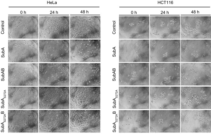
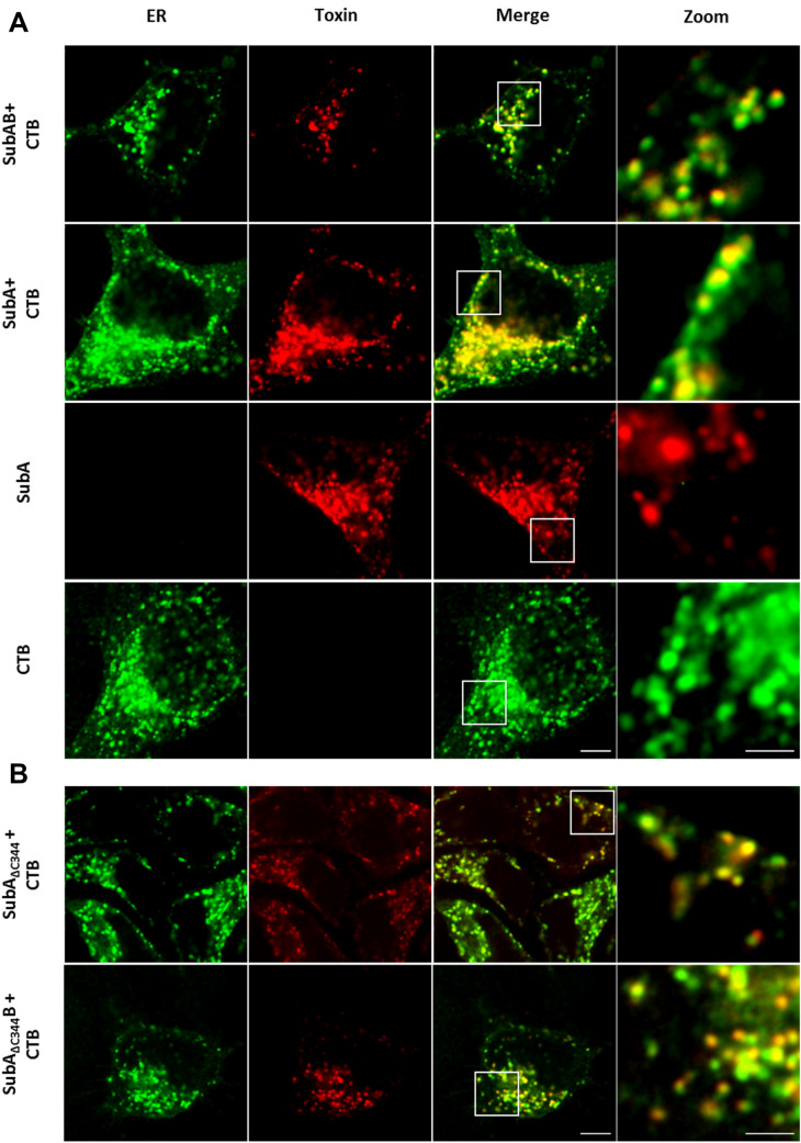
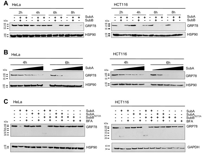
The subtilase cytotoxin (SubAB) is secreted by certain Shiga toxin-producing Escherichia coli (STEC) strains and is composed of the enzymatically active subunit SubA and the pentameric binding/transport subunit SubB. We previously demonstrated that SubA (10 µg/ml), in the absence of SubB, binds and intoxicates the human cervix cancer-derived epithelial cell line HeLa. However, the cellular and molecular mechanisms underlying the cytotoxic activity of SubA in the absence of SubB remained unclear. In the present study, the cytotoxic effects mediated by SubA alone were investigated in more detail in HeLa cells and the human colon cancer cell line HCT116. We found that in the absence of SubB, SubA (10 µg/ml) is internalized into the endoplasmic reticulum (ER), where it cleaves the chaperone GRP78, an already known substrate for SubA after its canonical uptake into cells via SubB. The autonomous cellular uptake of SubA and subsequent cleavage of GRP78 in cells is prevented by treatment of cells with 10 µM brefeldin A, which inhibits the transport of protein toxins into the ER. In addition, by analyzing the SubA mutant SubAΔC344, we identified the C-terminal SEEL motif as an ER-targeting signal. Conclusively, our results strongly suggest that SubA alone shares the same intracellular transport route and cytotoxic activity as the SubAB holotoxin.

Keywords: Cellular uptake; GRP78; Intracellular transport; Shiga toxin-producing Escherichia coli (STEC); Subtilase cytotoxin.


Conflict of interest statement

The authors declare no conflict of interest.

Figures

Fig. 1
Fig. 1
Cytotoxic effects of SubA2-2-His, SubAB2-2-His, SubA2-2-HisS272A and SubA2-2HisS272AB on HeLa and HCT116 cells. Cells were incubated with either 10 µg/ml of the single toxin component or a total protein concentration of 10 µg/ml for both toxin components in a molar ratio of 1:5 for 48 h at 37 °C. For control, cells were left untreated. To demonstrate changes in the cell morphology as well as in the cell number, images were taken after 0, 24, and 48 h. Experiments were performed in triplicates and representative images selected
Fig. 2
Fig. 2
a Confocal microscopic images of SubA2-2-His (red) co-localization with the ER (green) in HeLa cells. b Confocal microscopic images of SubA∆3442–2-His (red) co-localization with the ER (green) in HeLa cells. Cells were incubated for 6 h at 37 °C with 30 µg/ml ATTO647N-labeled SubA2-2-His (indicated as SubA), ATTO647N-labeled SubA∆3442–2-His (indicated as SubA∆344) or with 20 µg/ml SubAATTO-647NB2-2His (indicated as SubAB) or with SubA∆344,ATTO-647NB2-2His (indicated as SubA∆344B). The same cells were incubated with 15 µg/ml ALEXAFluor594-conjugated Cholera toxin B subunit (CTB) as established ER marker. In parallel, cells were only incubated with 30 µg/ml ATTO647N-labeled SubA2-2-His or ALEXAFluor594-conjugated CTB. Cells were then washed with PBS, fixed, blocked, and CTB (indicating localization of the ER) and the distribution of the labeled SubA2-2-His was captured via confocal microscopy. Scalebar: 5 µm (Zooms: 2 µm)
Fig. 3
Fig. 3
Western blot analysis of GRP78 cleavage due to SubA2-2-His treatment on HeLa and HCT116 cells. Cells, seeded in a 12-well plate, were incubated with or without toxin in FCS-free medium at 37 °C, as indicated. Cells were then solubilized in Lämmli sample buffer and subjected to SDS-PAGE. The substrate status was analyzed by Western blotting using monoclonal mouse anti GRP78 (see Methods). a Time course of GRP78 cleavage triggered by 5 µg/ml of either SubAB2-2-His (in a molar ratio of 1:5) or SubA2-2-His alone. b Time and concentration course of GRP78 cleavage. The following concentrations of SubA2-2-His, plotted from low to high, were used: 3.4 µg/ml, 6.8 µg/ml, 10.2 µg/ml, 13.6 µg/ml, and incubated for 4 h and 6 h. c Effect of brefeldin A (BFA) on GRP78 cleavage. Cells were pre-treated with or without 10 µM BFA and then treated with or without 10 µg/ml SubAB2-2-His (in a molar ratio of 1:5), SubAS272AB2-2-His, SubA2-2-His, SubAS272A2-2-His, or SubB2-2-His for 4 h at 37 °C
Fig. 4
Fig. 4
a Cytotoxic effects of 10 µg/ml SubAΔC3442-2-His or 10 µg/ml SubAΔC344B2-2-His (in a molar ratio of 1:5) on HeLa and HCT116 cells. Cells were incubated for 48 h at 37 °C. For control, cells were left untreated. To demonstrate changes in the cell morphology as well as in the cell number, images were taken after 0, 24, and 48 h. b, c Western blot analysis of GRP78 cleavage after treatment with SubAΔC3442-2-His. For b cells were seeded in 12-well plates and incubated with 10 µg/ml SubAΔC3442-2-His or a total protein concentration of 10 µg/ml (molar ratio of 1:5) for SubAΔC344B2-2-His in FCS-free medium for 4 h at 37 °C. For c cells were seeded in 12-well plates and incubated with increasing concentrations of SubA2-2-His and SubAΔC3442-2-His in FCS-free medium for 6 h at 37° C. The following concentrations of SubA2-2-His and SubAΔC3442-2-His, plotted from low to high, were used: HeLa cells: 2.5 µg/ml, 10 µg/ml, 20 µg/ml, 30 µg/ml, 40 µg/ml; HCT cells: 10 µg/ml, 20 µg/ml, 40 µg/ml. Cells were then solubilized in Lämmli sample buffer and subjected to SDS-PAGE. The substrate status, i.e. GRP78 cleavage, was analyzed by immunoblotting using monoclonal mouse anti GRP78 (see Methods)

Similar articles

Cited by

References

    1. Byres E, Paton AW, Paton JC, Löfling JC, Smith DF, Wilce MCJ, et al. Incorporation of a non-human glycan mediates human susceptibility to a bacterial toxin. Nature. 2008;456(7222):648–652. doi: 10.1038/nature07428. - DOI - PMC - PubMed
    1. de Castro E, Sigrist CJA, Gattiker A, Bulliard V, Langendijk-Genevaux PS, Gasteiger E, et al. ScanProsite: detection of PROSITE signature matches and ProRule-associated functional and structural residues in proteins. Nucleic Acids Res. 2006;34(Web server issue):W362–W365. doi: 10.1093/nar/gkl124. - DOI - PMC - PubMed
    1. Funk J, Biber N, Schneider M, Hauser E, Enzenmüller S, Förtsch C, et al. Cytotoxic and apoptotic effects of recombinant subtilase cytotoxin variants of Shiga toxin-producing Escherichia coli. Infect Immun. 2015;83(6):2338–2349. doi: 10.1128/IAI.00231-15. - DOI - PMC - PubMed
    1. Jerse AE, Yu J, Tall BD, Kaper JB. A genetic locus of enteropathogenic Escherichia coli necessary for the production of attaching and effacing lesions on tissue culture cells. Proc Natl Acad Sci USA. 1990;87(20):7839–7843. doi: 10.1073/pnas.87.20.7839. - DOI - PMC - PubMed
    1. Jores J, Rumer L, Wieler LH. Impact of the locus of enterocyte effacement pathogenicity island on the evolution of pathogenic Escherichia coli. Int J Med Microbiol. 2004;294(2–3):103–113. doi: 10.1016/j.ijmm.2004.06.024. - DOI - PubMed

Publication types

MeSH terms

LinkOut - more resources