. 2021 Apr;42(4):1255-1266.
doi: 10.1007/s10072-021-05068-7. Epub 2021 Jan 22.

Neuropathologic findings of patients with COVID-19: a systematic review

Affiliations

Neuropathologic findings of patients with COVID-19: a systematic review

Azalea T Pajo et al. Neurol Sci. 2021 Apr.

Abstract

Background: Despite the expanding literature that discusses insights into the clinical picture and mechanisms by which the SARS-CoV-2 virus invades the nervous system, data on the neuropathologic findings of patients who died following SARS-CoV-2 infection is limited.

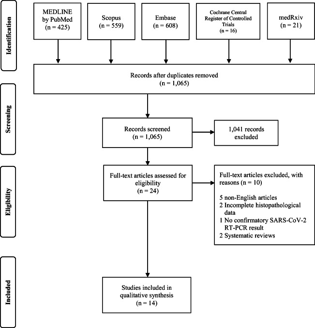
Methods: A broad literature search was done for published articles that reported on histopathological findings of the brain in patients with COVID-19 in PubMed by MEDLINE, Embase, CENTRAL by the Cochrane Library, and SCOPUS from December 31, 2019 to October 31, 2020.

Results: The systematic literature search strategy used resulted in a total of 1608 articles of which 14 were included in the analysis (PROSPERO registration number: CRD42020221022). There were ten case series, two case reports, one retrospective cohort, and one prospective cohort. The age of the patients ranged between 38 and 90 years old, most of them older than 65 years old (n=66, 45.2%) and males (n=79, 54.1%). Most tested negative in SARS-CoV-2 immunohistochemistry (n=70, 47.9%). The striking pathologic changes included diffuse edema (n=25, 17.1%), gliosis with diffuse activation of microglia and astrocytes (n=52, 35.6%), infarctions involving cortical and subcortical areas of the brain (n=4, 2.7%), intracranial bleed (subarachnoid hemorrhage and punctate hemorrhages) (n=18, 12.4%), arteriosclerosis (n=43, 29.5%), hypoxic-ischemic injury (n=41, 28.1%), and signs of inflammation (n=52, 35.6%). The cause of death was attributed to the cardiorespiratory system (n=66, 45.2%).

Conclusions: The neuropathologic changes observed likely represent direct cytopathic effects and indirect effects secondary to host-specific inflammatory response induced by the viral infection. Further studies however are required to better elucidate the pathologic mechanism.

Keywords: COVID-19; Neuropathology; SARS-CoV-2.


Conflict of interest statement

The authors declare no competing interests.

Figures

Fig. 1
Fig. 1
PRISMA flow diagram for study selection
Fig. 2
Fig. 2
Neuropathologic findings in patients with SARS-CoV-2 infection. At the level of the optic chiasm (A), there was a note of hypoxic changes, while at the level of the basal ganglia (B), prominent changes include watershed infarctions, hypoxic changes, punctate hemorrhages, and microbleeds. There was also a note of hypoxic and ischemic changes in the hippocampus, others with evidence of necrosis. Other pathologic changes observed include diffuse cerebral edema, gliosis with diffuse activation of microglia and astrocytes, infarctions involving cortical and subcortical areas of the brain, intracranial bleed (e.g., subarachnoid hemorrhage (SAH) and punctate hemorrhages), varying degrees of arteriosclerosis, evidence of hypoxic-ischemic injury, differing stages of inflammation as demonstrated by the presence of cellular components infiltrating into perivascular areas of the brain, presence of endovascular microthrombi, variable degrees of neuronal cell loss, and axonal degeneration/injury
Fig. 3
Fig. 3
Possible mechanisms underlying SARS-CoV-2-mediated neurological injury. SARS-CoV-2 can possibly gain access to the brain following three routes. (A) The virus could penetrate the CNS through the neuroepithelium of the olfactory mucosa to reach the olfactory bulb, gain access to the mitral cells and the olfactory nerve, and then spread to adjacent areas of the brain such as the hippocampus and other brain structures through retrograde viral transmission. (B and C) SARS-CoV-2 could also gain entry to the CNS by means of the vagus nerve through its terminals located along the respiratory and gastrointestinal tract. (D) Breach in the BBB where there is high expression of ACE2 receptors provides the mechanism for the hematogenous route of viral invasion

Similar articles

Cited by

References

    1. Tsivgoulis G, Palaiodimou L, Katsanos AH, Caso V, Köhrmann M, Molina C, Cordonnier C, Fischer U, Kelly P, Sharma VK, Chan AC, Zand R, Sarraj A, Schellinger PD, Voumvourakis KI, Grigoriadis N, Alexandrov AV, Tsiodras S. Neurological manifestations and implications of COVID-19 pandemic. Ther Adv Neurol Disord. 2020;13:175628642093203. doi: 10.1177/1756286420932036. - DOI - PMC - PubMed
    1. Wu Y, Xu X, Chen Z, Duan J, Hashimoto K, Yang L, Liu C, Yang C. Nervous system involvement after infection with COVID-19 and other coronaviruses. Brain Behav Immun. 2020;87:18–22. doi: 10.1016/j.bbi.2020.03.031. - DOI - PMC - PubMed
    1. Franca RA, Ugga L, Guadagno E, Russo D, Del Basso De Caro M. Neuroinvasive potential of SARS-CoV2 with neuroradiological and neuropathological findings: is the brain a target or a victim? APMIS. 2020;129:37–54. doi: 10.1111/apm.13092. - DOI - PubMed
    1. Collantes MEV, Espiritu AI, Sy MCC, Anlacan VMM, Jamora RDG. Neurological manifestations in COVID-19 infection: a systematic review and meta-analysis. Can J Neurol Sci. 2021;48:66–76. doi: 10.1017/cjn.2020.146. - DOI - PMC - PubMed
    1. Paniz-Mondolfi A, Bryce C, Grimes Z, Gordon RE, Reidy J, Lednicky J, Sordillo EM, Fowkes M. Central nervous system involvement by severe acute respiratory syndrome coronavirus-2 (SARS-CoV-2) J Med Virol. 2020;92:699–702. doi: 10.1002/jmv.25915. - DOI - PMC - PubMed

Publication types