Review
. 2021 Jun;46(6):2699-2711.
doi: 10.1007/s00261-020-02906-8. Epub 2021 Jan 23.

Bosniak classification of cystic renal masses, version 2019: interpretation pitfalls and recommendations to avoid misclassification

Affiliations
Review

Bosniak classification of cystic renal masses, version 2019: interpretation pitfalls and recommendations to avoid misclassification

Elizabeth Edney et al. Abdom Radiol (NY). 2021 Jun.

Abstract

The purpose of this review is to describe the potential sources of variability or discrepancy in interpretation of cystic renal masses under the Bosniak v2019 classification system. Strategies to avoid these pitfalls and clinical examples of diagnostic approaches are also presented. Potential pitfalls in the application of Bosniak v2019 are divided into three categories: interpretative, technical, and mass related. An organized, comprehensive review of possible discrepancies in interpreting Bosniak v2019 cystic masses is presented with pictorial examples of difficult clinical cases and proposed solutions. The scheme provided can guide readers to consistent, precise application of the classification system. Radiologists should be aware of the possible sources of misinterpretation of cystic renal masses when applying Bosniak v2019. Knowing which features and types of cystic masses are prone to interpretive errors, in addition to the inherent trade-offs between the CT and MR techniques used to characterize them, can help radiologists avoid these pitfalls.

Keywords: Cysts; Diagnostic error; Kidney; Kidney neoplasms; Magnetic resonance imaging; Renal cell carcinomas.


Figures

Fig. 1
Fig. 1
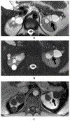
a – 67-year-old male with heterogeneous cystic renal mass at CT. The cystic mass appears mildly heterogeneous on portal venous phase CT (arrow). b – 67-year-old male with heterogeneous cystic renal mass at CT. Axial fat-saturated T1-weighted (T1W) contrast-enhanced spoiled gradient echo image confirms enhancement in the possible nodule (arrow), potentially leading to classification of this mass as Bosniak IV. c – 67-year-old male with heterogeneous cystic renal mass at CT. Coronal fat-saturated T1W contrast-enhanced spoiled gradient echo image demonstrates that the apparent nodule is not a nodule, but instead a confluence of minimally thickened (3 mm) septa (arrow). Note the horizontal orientation, which can mimic a nodule when viewed in the axial plane. Because the mass had thick septa (4 mm) elsewhere (not shown), it was characterized as Bosniak v2019 class III.
Fig. 2 –
Fig. 2 –
Challenges detecting enhancement at MRI when there is respiratory misregistration. Schematic T1-weighted MR images before and after contrast material administration (POST = post-contrast image; PRE = pre-contrast image; SUB = subtraction image [post-contrast minus pre-contrast]). Hemorrhagic or proteinaceous cystic masses contain intrinsically T1-hyperintense fluid (asterisk) that can conceal a nodule (solid arrow). In this example, an enhancing nodule is lower signal intensity than fluid on pre-contrast images (solid arrow) and isointense to fluid (i.e., invisible) on post-contrast images (dashed arrow). If the respiratory effort is sufficiently different between the pre- and post-contrast imaging, misregistration artifact will occur on subtraction imaging as bands of high and low signal intensity along the superior and inferior margins of the kidney and mass (arrowheads). Because the signal intensity of the enhancing nodule and hemorrhagic fluid is similar, mathematical subtraction of the signal intensity in the nodule minus the native high signal intensity of cyst fluid (i.e., due to misregistration) leads to obscuration of the enhancing nodule on subtraction imaging. Minimal residual signal intensity may be seen more inferiorly at the level of the nodule on subtraction images (open arrow). In this example, recognition of the hypointense pre-contrast nodule becoming isointense or invisible post-contrast allow a confident diagnosis of enhancement.
Fig. 3
Fig. 3
a – Discrepancies in evaluation of internal features in cystic renal masses on different sequences and modalities. Coronal T2W SSFSE image in a patient with a cystic renal mass with many (≥4) internal septa shows one to be 1.8 mm in thickness (arrow). b – Discrepancies in evaluation of internal features in cystic renal masses on different sequences and modalities. Coronal T1W contrast-enhanced spoiled gradient echo image in a patient with a cystic renal mass with many (≥4) septa. The same septum measures 3.0 mm. The image has some blurring. In this case, the discrepancy in measurements (1.8 mm on T2WI and 3.0 mm on post-contrast imaging) did not change the Bosniak v2019 class because many (≥4) thin (≤2 mm) septa and minimally thick (3 mm) septa are both features of a Bosniak IIF mass. c – Discrepancies in evaluation of internal features in cystic renal masses on different sequences and modalities. Axial T2W image in a different patient shows a cystic mass with many (≥4) internal septa. Apparent thickening of a septum > 4 mm (arrow) on this T2W image should be ignored because septa must enhance to be counted for Bosniak v2019 classes IIF, III, and IV. d – Discrepancies in evaluation of internal features in cystic renal masses on different sequences and modalities. Axial T1W contrast enhanced image shows the same septum measures <3 mm (arrow). Since Bosniak v2019 only allows enhancing septa to be counted, this was considered Bosniak IIF (many [≥4] smooth thin [≤2mm] enhancing septa). Although not directly addressed in Bosniak v2019, it is the opinion of the authors that all measurements should be rounded down to the nearest integer. e – Discrepancies in evaluation of internal features in cystic renal masses on different sequences and modalities. Axial contrast-enhanced CT shows a third patient with a cystic renal mass that appears homogeneous, nonenhancing, and without septa (Bosniak I). f – Discrepancies in evaluation of internal features in cystic renal masses on different sequences and modalities. Coronal T2W image of the same cystic mass as in (e) shows multiple thin septa (arrow). However, the septa did not enhance (not shown). Therefore, by Bosniak v2019, both the MRI and CT images are Bosniak v2019 class I cysts because septa must enhance to be counted.
Fig. 4
Fig. 4
a – 68-year-old male status post left nephrectomy for clear cell RCC. Unenhanced CT shows a mass in the posterior right kidney with attenuation of 15 HU. If homogeneous, it would be classified as Bosniak v2019 class II. However, after placing several small ROIs (Fig. 5b), the mass is shown to be heterogeneous. b – 68-year-old male status post left nephrectomy for clear cell RCC. Unenhanced CT with smaller, peripherally drawn ROIs demonstrate HU up to 25 HU. c – 68-year-old male status post left nephrectomy for clear cell RCC. Contrast-enhanced CT image shows enhancing tissue in > 25% of the mass (i.e., solid mass). The mass was pathologically proven to be clear cell RCC.
Fig. 5
Fig. 5
a – 73-year-old female with acute right upper quadrant pain. Coronal contrast-enhanced CT image reveals a heterogeneous, hyperdense mass abutting the right renal hilum without enhancement when compared to unenhanced CT images (not shown). Bosniak v2019 requires MRI to classify heterogenous non-enhancing masses at CT as these features could represent a papillary RCC. b – 73-year-old female with acute right upper quadrant pain. T2W single shot fast spin echo MR image performed 10 months later demonstrates marked decrease in size of what is now a mass with two components. The component abutting the hilum (short arrow) is low signal intensity. The exophytic component (long arrow) is high signal intensity. c – 73-year-old female with acute right upper quadrant pain. Precontrast fat-saturated T1W spoiled gradient echo image. The component abutting the hilum (short arrow) is high signal intensity consistent with hemorrhagic or proteinaceous fluid. The exophytic component (long arrow) is low signal intensity. d – 73-year-old female with acute right upper quadrant pain. Delayed postcontrast fat-saturated subtraction image demonstrates no enhancement in either component (arrows). Considering the marked decrease in size since prior imaging, this was likely an involuting hemorrhagic Bosniak II cyst.
Fig. 6
Fig. 6
a – 72-year-old male with abdominal pain and hematuria. Subtraction T1W spoiled gradient echo image during the nephrographic phase shows a large left renal mass with questionable central enhancement (arrow). b – 72-year-old male with abdominal pain and hematuria. Unenhanced T1W spoiled gradient echo image with an ROI drawn over the area in question shows a mean value of 73 signal intensity units. c – 72-year-old male with abdominal pain and hematuria. T1W spoiled gradient echo image during the nephrographic phase with an ROI drawn over the area in question shows a mean value of 91 signal intensity units, corresponding to a 23% increase in signal intensity compared to the unenhanced image (b). This mass was pathologically proven primitive neuroectodermal tumor (PNET) with extensive necrosis. Differentiating between a cystic mass and a largely necrotic solid mass may be challenging. The wall is minimally thickened and there is infiltrative enhancing tumor along the posterior margin (arrow), which are clues to the correct interpretation of a necrotic mass.
Fig. 7
Fig. 7
a – 67-year-old female with indeterminate renal mass on CT performed for evaluation of right upper quadrant pain. Axial T2W fat saturated SSFSE image demonstrates a T2 hyperintense right renal mass (white arrow), which could be mischaracterized as a Bosniak II cyst. Note, however, that the internal signal intensity does not match the level of intensity of the cerebrospinal fluid or the adjacent simple cyst. Additionally, the mass is slightly heterogeneous. b – 67-year-old female with indeterminate renal mass on CT performed for evaluation of right upper quadrant pain. Axial post contrast fat saturated T1W image shows heterogeneous enhancement of the mass (white arrow). Right partial nephrectomy confirmed clear cell RCC (Fuhrman grade 2). To assign a Bosniak II designation on the basis of T2WI alone, the mass must be homogeneous and as hyperintense as cerebrospinal fluid.
Fig. 8
Fig. 8
a – 25-year-old male with history of von Hippel-Lindau (VHL) undergoing follow-up. Axial T2W SSFSE image at baseline demonstrates multiple bilateral renal cysts. A left renal cyst (arrow) exhibits few (1 to 3) thin (≤2 mm) septa. Bosniak v2019 should not be applied to patients with a kidney cancer syndrome. b – 25-year-old male with history of VHL undergoing follow-up. Axial fat-saturated T2W SSFSE image performed approximately two years later shows enlargement of the solid component within the mass (arrow), now greater than 25% of the mass (i.e., now a solid mass). c – 25-year-old male with history of VHL undergoing follow-up. T1W spoiled gradient echo image during the nephrographic phase confirms solid enhancement (arrow). The mass was pathologically proven to be clear cell renal cell carcinoma.
Fig. 9
Fig. 9
a – 67-year-old female with bilateral incidental renal cysts on ultrasound. T1W fat saturated spoiled gradient echo image during the late nephrographic phase shows an enhancing nodule (compared to pre-contrast, not shown) within a superior left renal cystic mass (arrow) and a large homogeneous intermediate-signal intensity mass in the superior right kidney, which showed no enhancement (i.e. hemorrhagic/proteinaceous cyst). b – 67-year-old female with bilateral incidental renal cysts on ultrasound. Subtraction image of delayed postcontrast coronal T1W fat saturated spoiled gradient echo minus pre-contrast images performed 2 years later shows interval enlargement of the enhancing nodule with the left renal cystic mass (long arrow), diagnostic of Bosniak v2019 class IV. The superior right renal mass has decreased, consistent with involution of a Bosniak II hemorrhagic cyst, although the temporal change has created the appearance of enhancing minimally thick (3 mm) septa (short arrow) which could be misclassified as Bosniak IIF if the prior imaging were not available. The patient underwent left partial nephrectomy and clear cell papillary renal cell carcinoma was confirmed.
Fig. 10
Fig. 10
a – 58-year-old male with microhematuria. Contrast-enhanced CT image obtained during the excretory phase shows a cystic mass in the left kidney partially filling with excreted iodinated contrast (arrow), diagnostic of a calyceal diverticulum. b – 58-year-old male with microhematuria. Axial T1W gradient echo in phase image performed 2 years later demonstrates dependent stones (arrow). c – 58-year-old male with microhematuria. Subtraction image (delayed nephrographic minus precontrast) confirms enhancement of a solid nodule in the lateral aspect of the diverticulum (arrow), mimicking a Bosniak IV mass. However, a calyceal diverticulum should not be characterized with the Bosniak v2019 classification. Histopathologic analysis confirmed papillary urothelial carcinoma.

References

    1. Silverman SG, Pedrosa I, Ellis JH, et al. Bosniak Classification of Cystic Renal Masses, Version 2019: An Update Proposal and Needs Assessment. Radiology 2019; 292:475–488 - PMC - PubMed
    1. Davies L, Petitti DB, Woo M, Lin JS. Defining, Estimating, and Communicating Overdiagnosis in Cancer Screening. Ann Intern Med 2018; 169:739 - PubMed
    1. Esserman LJ, Thompson IM Jr., Reid B. Overdiagnosis and overtreatment in cancer: an opportunity for improvement. JAMA 2013; 310:797–798 - PubMed
    1. Tse JR, Shen J, Shen L, Yoon L, Kamaya A. Bosniak Classification of Cystic Renal Masses Version 2019: Comparison of Categorization using CT and MRI. AJR American journal of roentgenology 2020; - PubMed
    1. Tse JR, Shen J, Yoon L, Kamaya A. Bosniak Classification Version 2019 of Cystic Renal Masses Assessed With MRI. AJR American journal of roentgenology 2020; 215:413–419 - PubMed

Publication types

LinkOut - more resources