. 2021 Jan;1(1):e21.
doi: 10.1002/cpz1.21.

Differentiation of Brain Pericyte-Like Cells from Human Pluripotent Stem Cell-Derived Neural Crest

Affiliations

Differentiation of Brain Pericyte-Like Cells from Human Pluripotent Stem Cell-Derived Neural Crest

Benjamin D Gastfriend et al. Curr Protoc. 2021 Jan.

Erratum in

Abstract

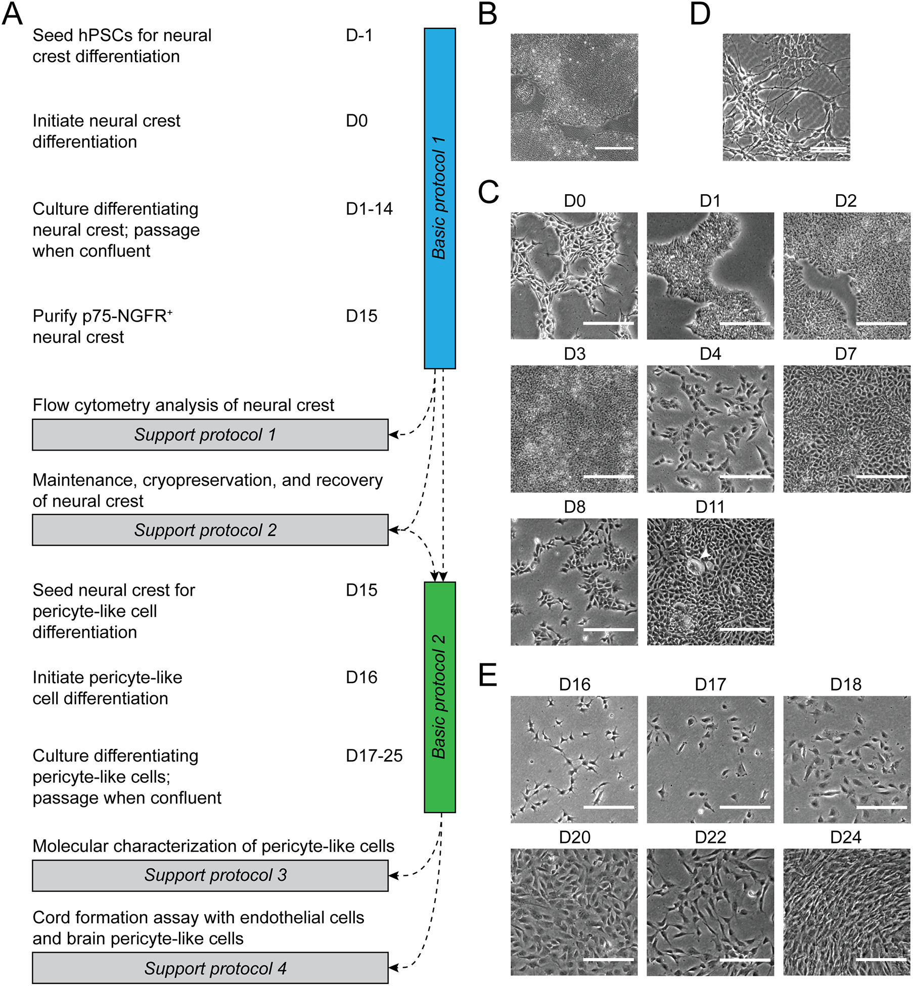
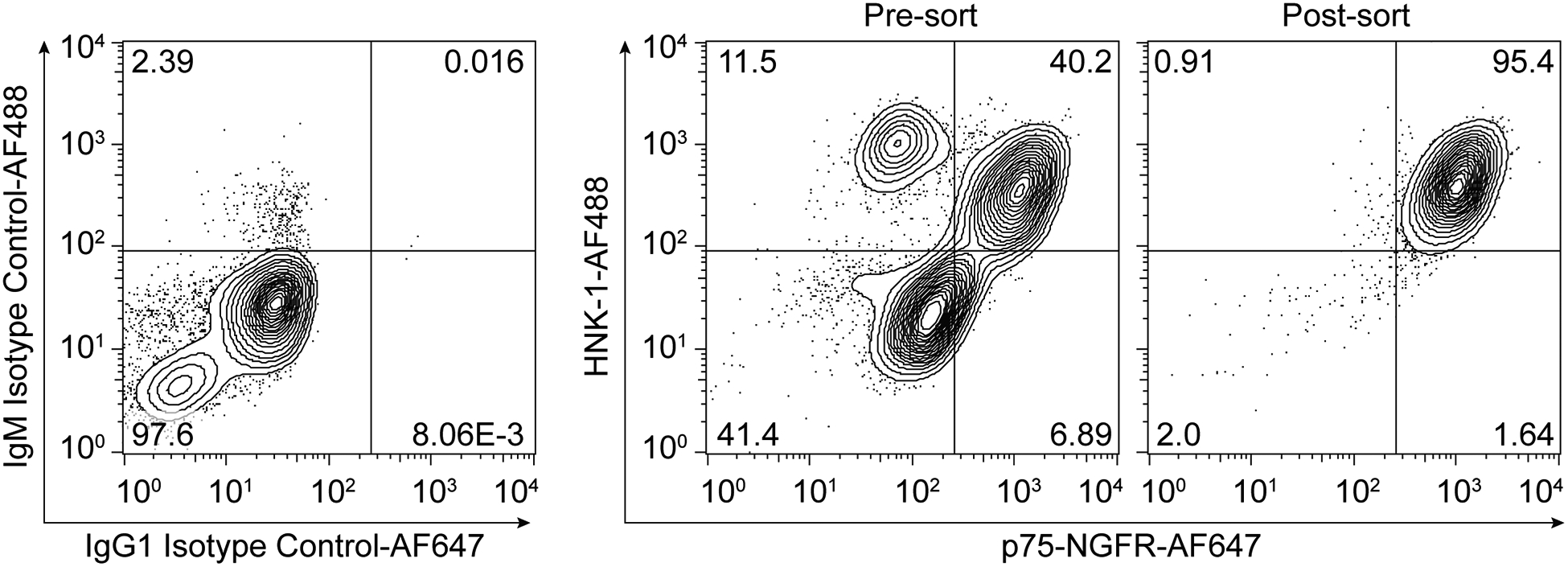
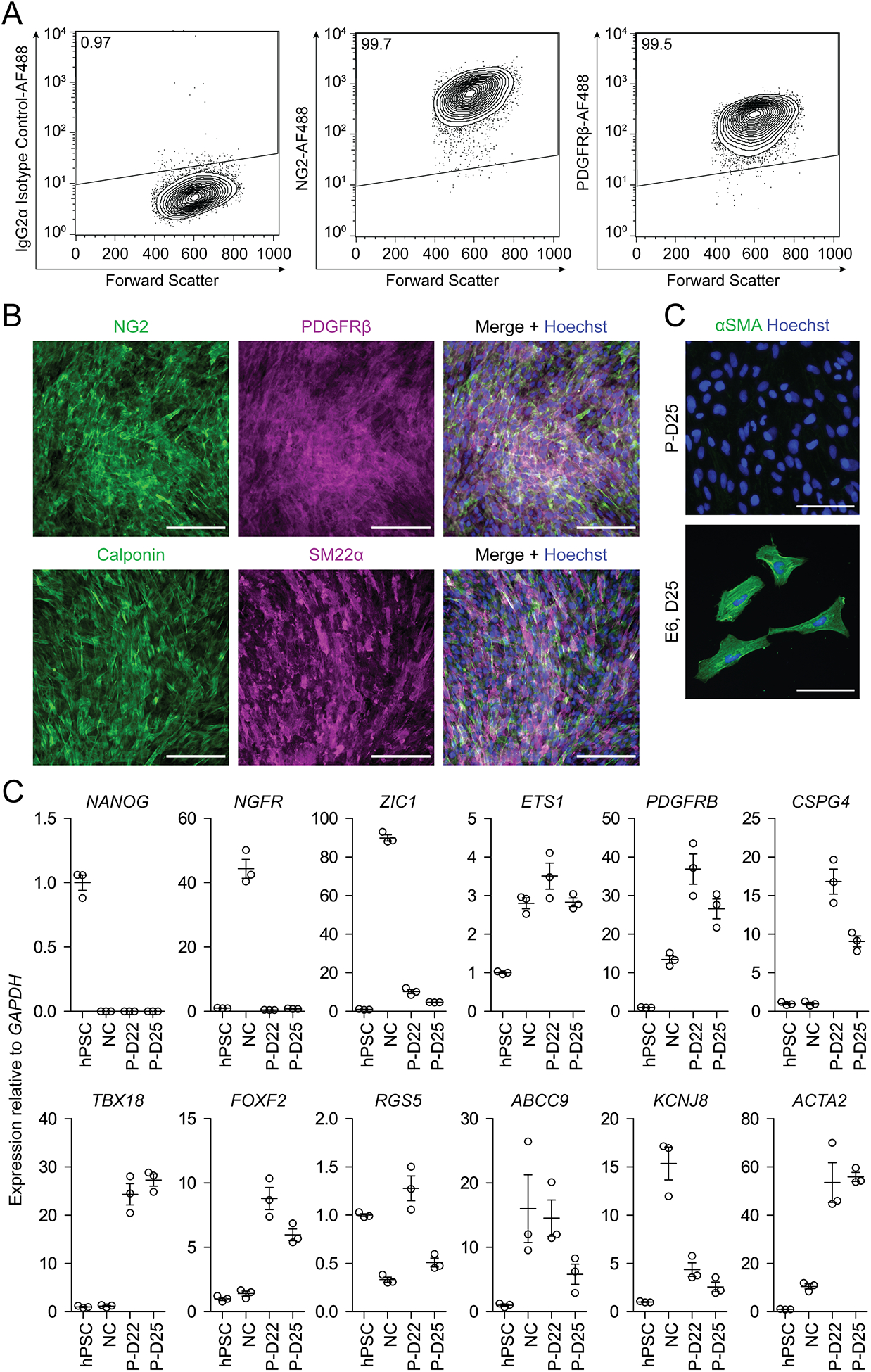
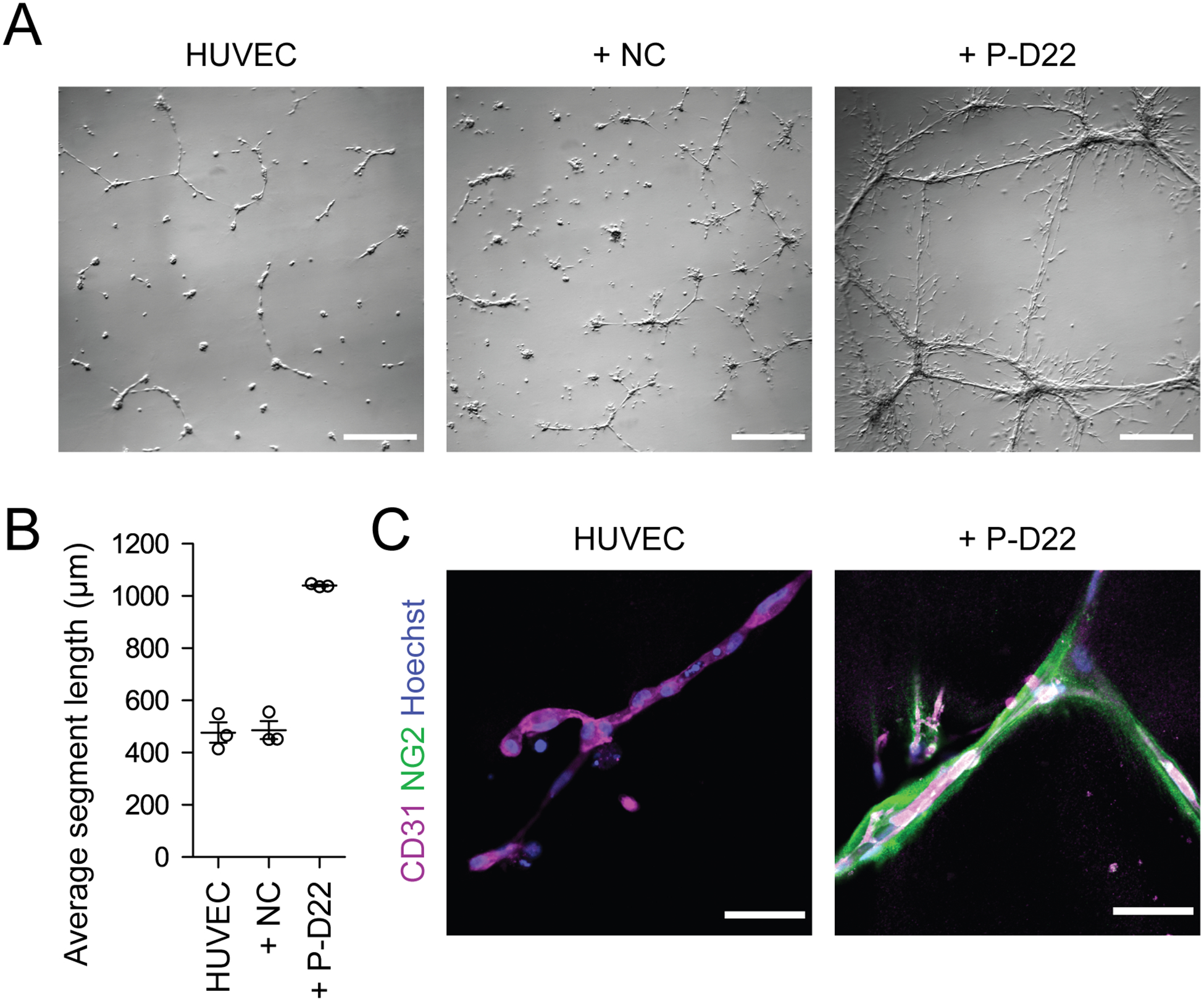
Brain pericytes regulate diverse aspects of neurovascular development and function, including blood-brain barrier (BBB) induction and maintenance. Primary brain pericytes have been widely employed in coculture-based in vitro models of the BBB, and a method to generate brain pericytes from human pluripotent stem cells (hPSCs) could provide a renewable, genetically tractable source of cells for BBB modeling and studying pericyte roles in development and disease. Here, we describe a protocol to differentiate hPSCs to NG2+ PDGFRβ+ αSMAlow brain pericyte-like cells in 22-25 days through a p75-NGFR+ HNK-1+ neural crest intermediate, which mimics the developmental origin of forebrain pericytes. The resulting brain pericyte-like cells have molecular and functional attributes of brain pericytes. We also provide protocols for maintenance, cryopreservation, and recovery of the neural crest intermediate, and for molecular and functional characterization of the resulting cells. © 2021 Wiley Periodicals LLC. Basic Protocol 1: Differentiation of hPSCs to neural crest Basic Protocol 2: Differentiation of neural crest to brain pericyte-like cells Support Protocol 1: Flow cytometry analysis of neural crest cells Support Protocol 2: Maintenance, cryopreservation, and recovery of neural crest cells Support Protocol 3: Molecular characterization of brain pericyte-like cells Support Protocol 4: Cord formation assay with endothelial cells and brain pericyte-like cells.

Keywords: blood-brain barrier; brain pericytes; human pluripotent stem cells; neural crest; neurovascular unit.


Conflict of interest statement

CONFLICT OF INTEREST

M.J.S, E.V.S, and S.P.P. are inventors on a patent application related to this work filed by the Wisconsin Alumni Research Foundation (US20200017827A1). The authors declare no other conflicts of interest.

Figures

Figure 1.
Figure 1.. Protocol overview.
(A) Schematic of protocol steps and timing. (B) Brightfield image of hPSC colony morphology at a density appropriate for seeding. Scale bar: 500 μm. (C) Brightfield images of typical cell morphology during the differentiation of hPSCs to neural crest. Arrowhead indicates a non-neural crest colony. Scale bars: 200 μm. (D) Example of spontaneous differentiation in a maintained neural crest culture. Scale bar: 100 μm. (E) Brightfield images of typical cell morphology during the differentiation of neural crest to brain pericyte-like cells. Scale bars: 200 μm.
Figure 2.
Figure 2.. Expected results of flow cytometry analysis of hPSC-derived neural crest.
Flow plots show isotype control staining and p75-NGFR/HNK-1 staining of hPSC-derived neural crest cells before MACS (pre-sort) and after two MACS steps (post-sort).
Figure 3.
Figure 3.. Expected results of molecular analysis of hPSC-derived brain pericyte-like cells.
(A) Flow cytometry analysis of D25 brain pericyte-like cells differentiated from hPSCs. Flow plots show isotype control staining, NG2 staining, and PDGFRβ staining. (B) Immunocytochemistry analysis of D25 brain pericyte-like cells stained for NG2/PDGFRβ and calponin/SM22⍺. Hoechst nuclear counterstain is overlaid in the merged images. Scale bars: 200 μm. (C) Immunocytochemistry analysis of D25 brain pericyte-like cells (P-D25) and neural crest-derived cells maintained in E6 medium from D16–D25 (E6, D25) stained for ⍺SMA. Cells in both conditions were passaged once, 1:2, upon reaching confluence. Hoechst nuclear counterstain is also shown. Scale bars: 100 μm. (D) qPCR analysis of hPSCs, neural crest (NC), brain pericyte-like cells at D22 (P-D22), and brain pericyte-like cells at D25 (P-D25). Expression of each gene of interest is shown relative to GAPDH expression and normalized to hPSC expression. Error bars represent the standard deviation of three replicate wells.
Figure 4.
Figure 4.. Expected results of cord formation assay.
(A) Phase contrast images of cells 24 h after initiating cord formation. Images of HUVECs alone, HUVECs cocultured with neural crest cells (+ NC), and HUVECs cocultured with brain pericyte like cells at D22 (+ P-D22) are shown. Scale bars: 1 mm. (B) Quantification of average segment length for the cord formation assay conditions described in (A). Error bars represent the standard deviation of three replicate chambers. (C) Confocal immunocytochemistry analysis of cords from HUVEC and + P-D22 conditions stained for CD31 and NG2. Hoechst nuclear counterstain is also shown. Scale bars: 50 μm.

References

    1. Alarcon-Martinez L, Yilmaz-Ozcan S, Yemisci M, Schallek J, Kılıç K, Can A, Di Polo A, and Dalkara T 2018. Capillary pericytes express α-smooth muscle actin, which requires prevention of filamentous-actin depolymerization for detection. eLife 7:1–17. - PMC - PubMed
    1. Ando K, Fukuhara S, Izumi N, Nakajima H, Fukui H, Kelsh RN, and Mochizuki N 2016. Clarification of mural cell coverage of vascular endothelial cells by live imaging of zebrafish. Development (Cambridge) 143:1328–1339. - PMC - PubMed
    1. Armulik A, Genové G, and Betsholtz C 2011. Pericytes: Developmental, Physiological, and Pathological Perspectives, Problems, and Promises. Developmental Cell 21:193–215. Available at: https://linkinghub.elsevier.com/retrieve/pii/S1534580711002693. - PubMed
    1. Armulik A, Genové G, Mäe M, Nisancioglu MH, Wallgard E, Niaudet C, He L, Norlin J, Lindblom P, Strittmatter K, et al. 2010. Pericytes regulate the blood-brain barrier. Nature 468:557–561. - PubMed
    1. Bell RD, Winkler EA, Sagare AP, Singh I, LaRue B, Deane R, and Zlokovic BV 2010. Pericytes Control Key Neurovascular Functions and Neuronal Phenotype in the Adult Brain and during Brain Aging. Neuron 68:409–427. Available at: 10.1016/j.neuron.2010.09.043. - DOI - PMC - PubMed

LinkOut - more resources