Review
. 2021 Jul:174:87-113.
doi: 10.1016/j.addr.2021.01.011. Epub 2021 Jan 21.

Modulating the foreign body response of implants for diabetes treatment

Affiliations
Review

Modulating the foreign body response of implants for diabetes treatment

Bhushan N Kharbikar et al. Adv Drug Deliv Rev. 2021 Jul.

Abstract

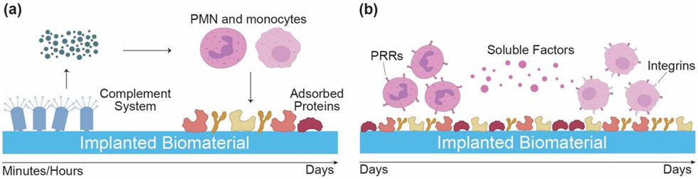
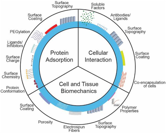
Diabetes Mellitus is a group of diseases characterized by high blood glucose levels due to patients' inability to produce sufficient insulin. Current interventions often require implants that can detect and correct high blood glucose levels with minimal patient intervention. However, these implantable technologies have not reached their full potential in vivo due to the foreign body response and subsequent development of fibrosis. Therefore, for long-term function of implants, modulating the initial immune response is crucial in preventing the activation and progression of the immune cascade. This review discusses the different molecular mechanisms and cellular interactions involved in the activation and progression of foreign body response (FBR) and fibrosis, specifically for implants used in diabetes. We also highlight the various strategies and techniques that have been used for immunomodulation and prevention of fibrosis. We investigate how these general strategies have been applied to implants used for the treatment of diabetes, offering insights on how these devices can be further modified to circumvent FBR and fibrosis.

Keywords: Biomaterials; Diabetes; Encapsulation; Fibrosis; Foreign body response; Immune system; Implants; Sensors; Type I diabetes; Type II diabetes.


Figures

Figure 1.
Figure 1.
Multiple interrelated pathways activate the immune cascade post-implantation. (a) Soluble factors released from the activation of complement system (C3a, C5a), prime polymorphonuclear leukocytes (PMN) and macrophages. (b) The primed immune cells interact with adsorbed proteins through pattern recognition receptors (PRRs) that recognize pattern associated molecular patterns (PAMPs) on biomaterial. Soluble factors released from PMNs further activate monocytes, which use both PRR and integrins to interact with the implanted biomaterial. Monocytes differentiate into macrophages and control the subsequent immune response.
Figure 2.
Figure 2.
The fate of the implant depends the resolution of the inflammatory immune cascade. (a) If macrophages are able to polarize from the inflammatory stage (M1) to their reparative stage (M2), they release soluble factors that promote fibroblasts to secrete collagen and promote integration of the implant. (b) If macrophages are unable to successfully transition from M1 to M2 phenotype, foreign body giant cells (FBGC) form and adhere to the implant surface. FBGC secrete more inflammatory soluble factors that activate myofibroblasts (fibrotic phenotype of fibroblasts), which secrete excessive amounts of collagen, leading to fibrous encapsulation of implant.
Figure 3.
Figure 3.
Macrophages and dendritic cells work together to activate the adaptive immune system. Dendritic cells and sometimes, macrophages present antigens to T cells that stimulate activation of different T cell subtypes. These subtypes are influenced by the soluble factors present in the local microenvironment. If pro-inflammatory macrophages are present, the secreted soluble factors activate the inflammatory Th1 CD4 cells. Meanwhile, if reparative macrophages are present, they secrete factors that activate the reparative Th2 CD4 cells along with regulatory CD4 (Treg) cells.
Figure 4.
Figure 4.
Several strategies can be used to mitigate the FBR and resulting fibrotic overgrowth. The three major categories that are affected by changes in material properties are protein adsorption, cell and tissue biomechanics, and cellular interaction. Changes in any of these three categories can induce a favorable immune response towards implants and increase their longevity as well as function in vivo.
Figure 5.
Figure 5.
Chitosan-coated alginate capsules show demonstrate high efficacy and biocompatibility as islet microencapsulation vehicles. (a) After 1 year of xenogenic islet transplantation, Masson’s trichrome staining chitosan-alginate capsules show lower pericapsular fibrillar collagen network as compared to alginate capsules. (b) Oral blood glucose tolerance test of diabetic canines with 1 year of allogenic islet transplantation showed similar results to test conducted on non-diabetic canines. (c) Moreover, there was no gross fibrosis or cell adhesion on any of the 3 beagles after 1 year of implantation, highlighting the biocompatibility of these capsules [380].
Figure 6.
Figure 6.
Long-term delivery of drugs in compact crystals can help modulate fibrosis caused by implantation of alginate microcapsules (a) FACs analysis shows that several drugs are able to modulate the effects of alginate microcapsules on recruiting macrophages and neutrophils (b) H&E and Masson Trichrome staining shows that, after 3 months, several crystalline drugs are able to reduce fibrosis typically induced by alginate spheres in the subcutaneous space. (c) In particular, co-encapsulation of crystalline GW250 with alginate microspheres showed little to no fibrotic overgrowth even after 6 months of implantation in the omentum or subcutaneous space. (d) Moreover, transplantation of islets co-encapsulated with crystalline GW250 in diabetic mice are able to maintain euglycemia for a prolonged period as compared to no drug containing microcapsules and amorphous-loaded microcapsules. (e) Photos also showed high collagenous deposition around control capsules with no drug as compared to capsules with crystalline drug after 72 days of transplant in the subcutaneous space [390].
Figure 7.
Figure 7.
Calcium releasing twist-folded nylon sutures which allow in situ crosslinking method of alginate hydrogel have great potential as islet macroencapsulation devices. (a) This thread-reinforced alginate fiber for islet encapsulation (TRAFFIC) device offers a new technology that combines polymeric and hydrogel approach to encapsulate cells. (b) Moreover, the easily modifiable devices showed little to no cellular overgrowth and correction of diabetes in rats after 7-month of implantation in the intraperitoneal cavity. When tested in dogs, not only did the scientists show that this device is easily scalable but histological evidence also showed high biocompatibility, demonstrating the translatability of this device.
Figure 8.
Figure 8.
Relevant ancillary approaches for mitigating foreign body response and fibrosis which can be extended to the implants for the diabetes. (a) An implantable and actuatable soft reservoir device modulates host foreign body response and describe milli-scale DSRs that successfully use a mechanoceutical approach to actively modulate the biomechanics of the biotic-abiotic interface by perturbing strain and fluid flow [462]. (b) A wireless resonant magnetoelastic actuators made from metal alloy foils generate a fluid flow on the surfaces of implantable Ahmed glaucoma drainage devices to limit cellular adhesion to the surface of the implant to prevent encapsulation and failure [463]. (c) A micro-engineered non-resorbable nano-patterned biosynthesized cellulose (BC) membrane as conformal wrapping around cardiac implantable electronic device (CIED) creating BC interface between the implant and the surrounding tissue in the surgical pocket which significantly reduce the formation of fibrotic tissue [464]. (d) The crystallized drug with compact lattice structure for the long-term controlled release of drug for local anti-inflammatory effect for the suppression of foreign body reaction [371].

Similar articles

Cited by

References

    1. Jiménez PG, Martín-Carmona J, Hernández EL, Diabetes mellitus, Med. (2020). 10.1016/j.med.2020.09.010. - DOI
    1. Atkinson MA, Eisenbarth GS, Michels AW, Type 1 diabetes, Lancet. (2014). 10.1016/S0140-6736(13)60591-7. - DOI - PMC - PubMed
    1. Katsarou A, Gudbjörnsdottir S, Rawshani A, Dabelea D, Bonifacio E, Anderson BJ, Jacobsen LM, Schatz DA, Lernmark A, Type 1 diabetes mellitus, Nat. Rev. Dis. Prim (2017). 10.1038/nrdp.2017.16. - DOI - PubMed
    1. Paschou SA, Papadopoulou-Marketou N, Chrousos GP, Kanaka-Gantenbein C, On type 1 diabetes mellitus pathogenesis, Endocr. Connect (2018). 10.1530/EC-17-0347. - DOI - PMC - PubMed
    1. Samson SL, Garber AJ, Type 2 diabetes, in: Encycl. Endocr. Dis, 2018. 10.1016/B978-0-12-801238-3.95795-7. - DOI

Publication types