Review
. 2021 Mar:136:109548.
doi: 10.1016/j.ejrad.2021.109548. Epub 2021 Jan 14.

Radiological, epidemiological and clinical patterns of pulmonary viral infections

Affiliations
Review

Radiological, epidemiological and clinical patterns of pulmonary viral infections

Konstantinos Stefanidis et al. Eur J Radiol. 2021 Mar.

Abstract

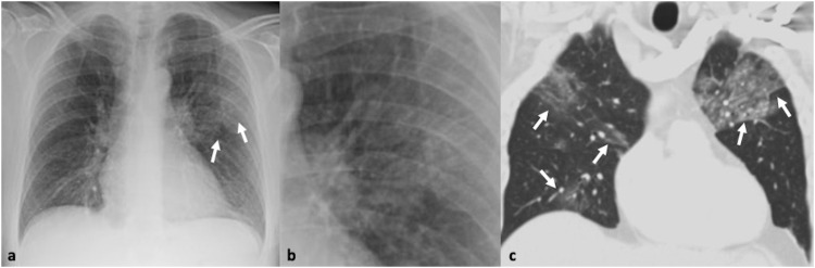
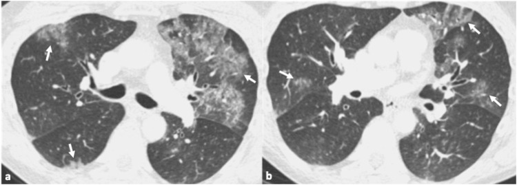
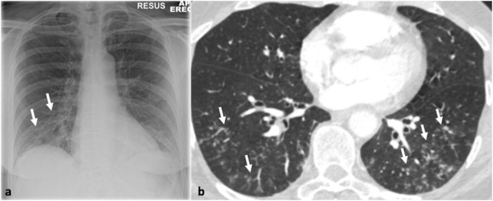
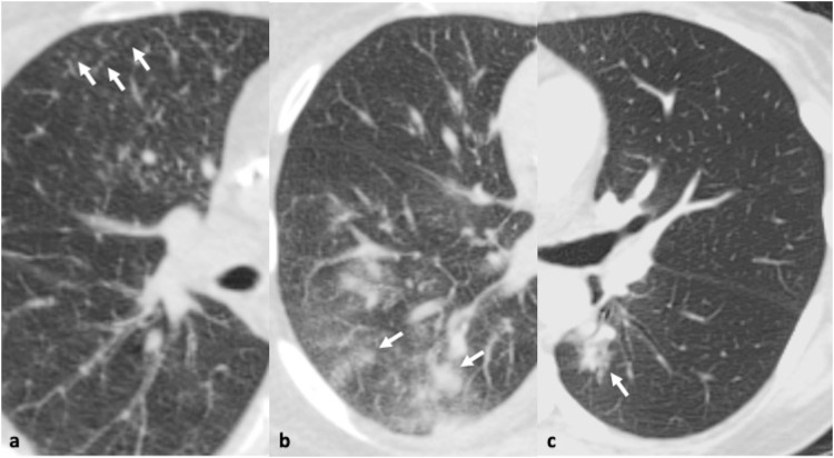
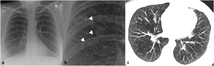
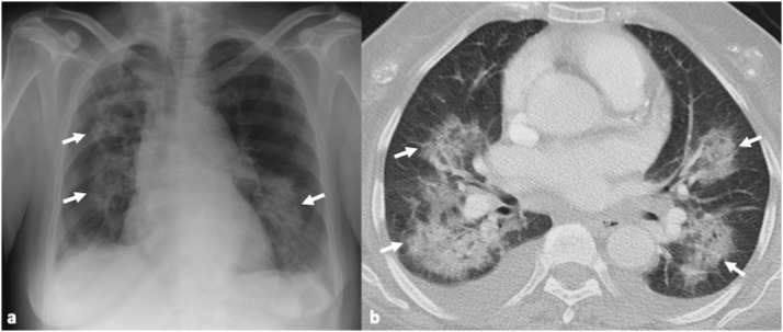
Respiratory viruses are the most common causes of acute respiratory infections. However, identification of the underlying viral pathogen may not always be easy. Clinical presentations of respiratory viral infections usually overlap and may mimic those of diseases caused by bacteria. However, certain imaging morphologic patterns may suggest a particular viral pathogen as the cause of the infection. Although definitive diagnosis cannot be made on the basis of clinical or imaging features alone, the use of a combination of clinical and radiographic findings can substantially improve the accuracy of diagnosis. The purpose of this review is to present the clinical, epidemiological and radiological patterns of lower respiratory tract viral pathogens providing a comprehensive approach for their diagnosis and identification in hospitals and community outbreaks.

Keywords: Chest x-ray; Clinical presentation; Computed Tomography; Diagnosis; Differential diagnosis; Epidemiology; Imaging; Respiratory viruses.


Conflict of interest statement

The authors report no declarations of interest.

Figures

Fig. 1
Fig. 1
41-year-old male with confirmed influenza pneumonia who presented with fever, dry cough and myalgia. 1a, b. Chest x-ray and magnified view shows left upper lobe patchy increased air-space opacification (arrows). 1c. Coronal CT image confirms the left upper lobe ground glass opacity and further multifocal ground glass involvement in both lungs (arrows).
Fig. 2
Fig. 2
45-year-old male patient with confirmed influenza pneumonia. 2a, b. Axial CT images show multifocal patchy ground glass opacities (arrows).
Fig. 3
Fig. 3
54-year-old woman with fever and cough. Parainfluenza was the only pathogen recovered from respiratory secretions. 3a. Chest x-ray shows reticulonodular opacities in the right lower zone (arrows). 3b. Transverse thin section CT scan confirms the presence of multiple micronodules and branching opacities (arrows).
Fig. 4
Fig. 4
51-year-old woman with confirmed respiratory syncytial virus infection. 4a-c. Transverse thin section CT scan through the mid and lower zones demonstrates a few tiny nodules (arrows, a) and bilateral patchy areas of nodular ground glass opacity and consolidation (arrows b, c).
Fig. 5
Fig. 5
64-year-old male with confirmed rhinovirus infection who presented with sore throat and cough with. 5a-c. Axial thin section CT scan demonstrates a few nodules (arrowheads, a, b) and areas of ground glass opacity (arrows, c) in the right lower lobe. Bronchial wall thickening and mild mosaicism is also noted in the left lung base, corroborating small airway inflammation.
Fig. 6
Fig. 6
47-year-old male with pneumonia due to human metapneumovirus. 6a-c. Axial CT images in the right apical region and right and left mid zone reveal three coexisting patterns: ground glass opacity (arrows, a-c), reticular changes and ill-defined centrilobular nodules (arrowheads, c).
Fig. 7
Fig. 7
31-year-old male with pneumonia due to MERS coronavirus who presented with cough and sputum. 7a-c. Axial and coronal CT images show bilateral symmetrical ground glass opacity with an upper and mid zone distribution. 7d. Small field of view in the right upper lobe at the level of the carina demonstrates a crazy-paving pattern.
Fig. 8
Fig. 8
61-year-old male with confirmed Covid-19 pneumonia. Series of chest x-rays show the progressive radiographic changes. 8a. Peripheral distribution of the airspace opacities in day 5 of admission (arrows, a). 8b, c. Radiographic progression with diffuse bilateral involvement of the lungs in day 8 (b) and 10 of admission (c).
Fig. 9
Fig. 9
51-year-old male with confirmed Covid-19 pneumonia. 9a, b. Axial CT images show typical CT appearances with bilateral peripheral ground glass opacities (arrows).
Fig. 10
Fig. 10
59-year-old male with confirmed Covid-19 pneumonia. Axial CT image shows nodular round ground glass discrete opacities bilaterally (arrows).
Fig. 11
Fig. 11
59-year-old man with confirmed Covid-19 pneumonia who presented with chest pain, cough, and fever. 11a. Axial CT image shows ground glass opacity and a crazy-paving pattern with peripheral distribution (arrows). 11b. In the same patient, axial CT image 5 days later shows at the same level, progression to organizing consolidation (arrows) and some fibrotic bands indicative of the late peak and early absorption stage.
Fig. 12
Fig. 12
42-year-old male with fever following bone marrow transplantation. Adenovirus was the only pathogen recovered from respiratory secretions. Axial CT image shows segmental consolidation with mild surrounding ground glass in the right lower lobe (arrows). A small subpleural focus of consolidation is also seen.
Fig. 13
Fig. 13
35-year-old male with healed varicella-zoster infection. 13a, b. Chest x-ray and close-up view shows multiple, bilateral and randomly distributed tiny calcified nodules (arrowheads). 13c. Axial CT image confirms the findings of the chest radiograph.
Fig. 14
Fig. 14
67-year-old female patient with pneumonia due to cytomegalovirus following kidney transplantation. 14a. Chest x-ray shows bilateral air-space opacification (arrows). 14b. Axial CT image shows the bilateral patchy areas of consolidation and ground glass opacity (arrows) associated with bilateral pleural effusions.

References

    1. Fears J.R. The plague under Marcus Aurelius and the decline and fall of the Roman Empire. Infect. Dis. Clin. North Am. 2004;18(March (1)):65–77. doi: 10.1016/s0891-5520(03)00089-8. - DOI - PubMed
    1. Suzuki A. Smallpox and the epidemiological heritage of modern Japan: towards a total history. Med. Hist. 2011;55(July (3)):313–318. doi: 10.1017/s0025727300005329. - DOI - PMC - PubMed
    1. Centers for Disease Control and Prevention, Past pandemics, Available at: https://www.cdc.gov/flu/pandemic-resources/basics/past-pandemics.html (Accessed 09 October 2020).
    1. Kobasa D., Kawaoka Y. Emerging influenza viruses: past and present. Curr. Mol. Med. 2005;5(December (8)):791–803. doi: 10.2174/156652405774962281. - DOI - PubMed
    1. Corman V.M., Muth D., Niemeyer D., Drosten C. Hosts and sources of endemic human coronaviruses. Adv. Virus Res. 2018;100:163–188. doi: 10.1016/bs.aivir.2018.01.001. - DOI - PMC - PubMed