. 2021 Jan 23;14(1):29.
doi: 10.1186/s13068-020-01860-3.

Four cellulose-active lytic polysaccharide monooxygenases from Cellulomonas species

Affiliations

Four cellulose-active lytic polysaccharide monooxygenases from Cellulomonas species

James Li et al. Biotechnol Biofuels. .

Abstract

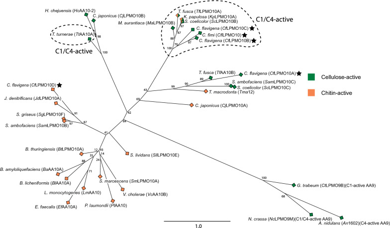
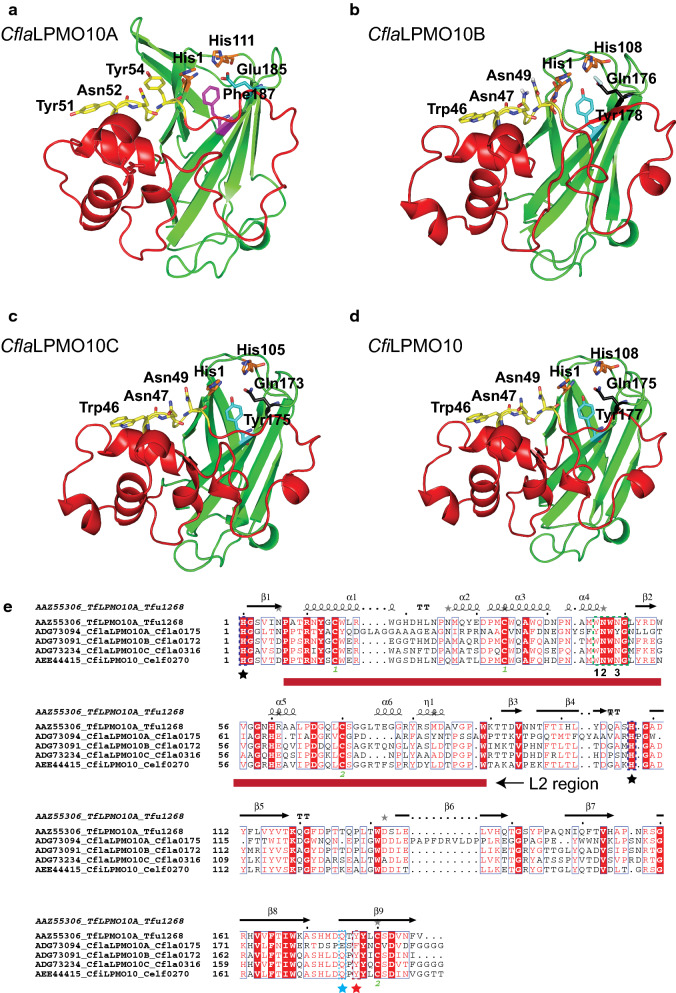
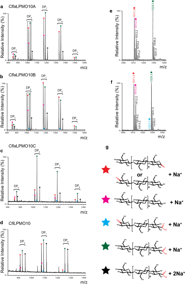
Background: The discovery of lytic polysaccharide monooxygenases (LPMOs) has fundamentally changed our understanding of microbial lignocellulose degradation. Cellulomonas bacteria have a rich history of study due to their ability to degrade recalcitrant cellulose, yet little is known about the predicted LPMOs that they encode from Auxiliary Activity Family 10 (AA10).

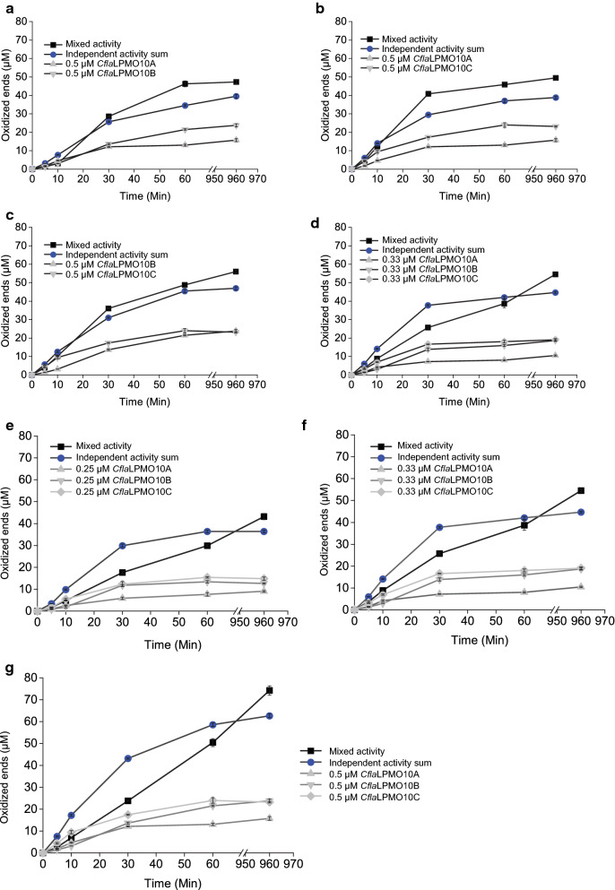
Results: Here, we present the comprehensive biochemical characterization of three AA10 LPMOs from Cellulomonas flavigena (CflaLPMO10A, CflaLPMO10B, and CflaLPMO10C) and one LPMO from Cellulomonas fimi (CfiLPMO10). We demonstrate that these four enzymes oxidize insoluble cellulose with C1 regioselectivity and show a preference for substrates with high surface area. In addition, CflaLPMO10B, CflaLPMO10C, and CfiLPMO10 exhibit limited capacity to perform mixed C1/C4 regioselective oxidative cleavage. Thermostability analysis indicates that these LPMOs can refold spontaneously following denaturation dependent on the presence of copper coordination. Scanning and transmission electron microscopy revealed substrate-specific surface and structural morphological changes following LPMO action on Avicel and phosphoric acid-swollen cellulose (PASC). Further, we demonstrate that the LPMOs encoded by Cellulomonas flavigena exhibit synergy in cellulose degradation, which is due in part to decreased autoinactivation.

Conclusions: Together, these results advance understanding of the cellulose utilization machinery of historically important Cellulomonas species beyond hydrolytic enzymes to include lytic cleavage. This work also contributes to the broader mapping of enzyme activity in Auxiliary Activity Family 10 and provides new biocatalysts for potential applications in biomass modification.

Keywords: AA10; Bioethanol; Biomass; Bioproducts; Cellulose; LPMO.


Conflict of interest statement

The authors declare that they have no competing interests.

Figures

Fig. 1
Fig. 1
Maximum-likelihood phylogenetic tree of all currently characterized AA10 LPMO catalytic modules. Full-length protein sequences were retrieved from GenBank via the CAZy database and manually truncated to remove the signal peptide and C-terminal domains (guided in part by SignalP and BLASTP analysis). The catalytic sequences were then aligned using MUSCLE and manually refined to preserve the position of the catalytic residues prior to maximum-likelihood analysis using RAxML 8.2.10 utilizing a JTT matrix-based nucleotide substitution model and automatic halting bootstraps (702 bootstraps). Three characterized AA9 LPMO catalytic module sequences with different regioselective activities on chitin were used as outgroups (GtLPMO9B [131], AN1602 [132], and NcLPMO9D [133]). The input and output files are provided as Additional file1: S1 and Additional file2: S2
Fig. 2
Fig. 2
Homology models and primary sequence alignment of Cellulomonas LPMO catalytic modules. ad Cartoon representation of three-dimensional homology models of CflaLPMO10A, CflaLPMO10B, CflaLPMO10C, and CfiLPMO10, respectively, generated using the Phyre2 server with T. fusca LPMO10A as the template structure [81]. The active-site histidine residues are depicted as orange sticks, the three conserved residues of the cellulose-active motif are depicted as yellow sticks, the axial tyrosine or phenylalanine residues are depicted as turquoise or pink sticks, respectively, and the catalytic glutamate or glutamine residues are depicted as teal sticks. Residue numbering corresponds to His-1 in the mature protein sequence. The L2 region is colored red to distinguish it from the immunoglobulin-like β-sandwich core colored in green. e Sequence and secondary structure alignment of Cellulomonas LPMOs compared to T. fusca LPMO10A [73]. The cellulose-active motif is indicated by a green dotted box and the positioning of the conserved residues mentioned in-text are numbered 1–3. The two active site histidine residues are indicated with black star symbols. The red dotted box with star indicates the position of the axial aromatic residue. The position of the catalytic glutamate or glutamine is indicated with a teal star
Fig. 3
Fig. 3
HPAEC-PAD analysis of Cellulomonas LPMO activity on PASC. a Chromatogram over the retention times 3–25 min b Expansion of the 16–24 min region. Native and C1-oxidized cello-oligosaccharide standard peaks are annotated. All activity assays were conducted using 1 μM of enzyme, 0.1% PASC, and 1 mM ascorbic acid over 16 h at 37 °C. The negative control (NoEnz Ctrl) contained all assay components except enzyme
Fig. 4
Fig. 4
MALDI-TOF product analysis of Cellulomonas LPMO activity on PASC. ad Mass spectrum of m/z 800–1600 showing native and oxidized cello-oligosaccharide products between DP5—DP8 [M + Na+] released by the Cellulomonas LPMO indicated above each respective panel. e, f Representative expanded mass spectra of m/z 1000–1040 depicting DP6 [M + Na+] peaks from CflaLPMO10A and CflaLPMO10B corresponding to native, C1-, and mixed C1/C4-oxidized cello-oligosaccharides. g Symbols used to denote native and oxidized cello-oligosaccharide products corresponding to peaks in mass spectra. Red stars are monosodiated aldonic and lactone forms of C1-oxidized cello-oligosaccharides. Pink stars are monosodiated native cello-oligosaccharides; teal stars are monosodiated doubly oxidized C1 aldonic acid/C4 ketoaldose oligosaccharides; green stars are monosodiated aldonic acid forms of C1-oxidized cello-oligosaccharides; and black stars are disodiated aldonic acid forms of C1-oxidized cello-oligosaccharides
Fig. 5
Fig. 5
C1-oxidized ends produced by 1 μM Cellulomonas LPMOs after 16 h on a panel of insoluble cellulose substrates. All substrates were assayed at 0.1% except NBSKP fiber which was assayed at 1% (no activity was detected at 0.1% NBSKP fiber). For each substrate, T. reesei cellulase cocktail (Celluclast; Sigma-Aldrich, P/N: C2730-30) was used to convert all C1-oxidized products into cellobionic acid and quantified as the total C1-oxidized ends generated (μM). Total oxidized ends were obtained by quantifying cellobionic acid by HPLC following hydrolysis of soluble products with T. reesei Celluclast enzyme cocktail. Each time point represents the average of three independent assays measured singly by HPLC, with error bars indicating the standard error of the mean
Fig. 6
Fig. 6
Surface morphology of Cellulomonas LPMO-oxidized cellulose visualized under scanning and transmission electron microscopy. a SEM of untreated (control) and LPMO-oxidized Avicel. b TEM of untreated (control) and LPMO-oxidized Avicel. c SEM of untreated (control) and LPMO-oxidized PASC. d TEM of untreated (control) and LPMO-oxidized PASC. Scale bars and magnification values are indicated below each representative micrograph
Fig. 7
Fig. 7
Progress curves of released soluble oxidized products by C. flavigena LPMOs incubated in combination and independently. ad Double and triple combinations of C. flavigena enzymes incubated together at a total enzyme loading of 1 μM compared to the sum total of the independent enzyme proportions at 1 μM. eg Boosting effect of a combined mixture of all three C. flavigena LPMOs at three different enzyme loading concentrations vs the sum total of independent enzyme proportions at each respective total enzyme load. Total oxidative activity was quantified from simplification of the product profile to cellobionic acid using T. reesei cellulase cocktail as described in the results and each assay was performed independently and in triplicate to calculate standard error of the mean

References

    1. Bidlack J, Malone M, Benson R. Molecular structure and component integration of secondary cell walls in plants. Proc Oklahoma Acad Sci. 1992;72:51–56.
    1. Hanus J, Mazeau K. The xyloglucan–cellulose assembly at the atomic scale. Biopolymers. 2006;82(1):59–73. doi: 10.1002/bip.20460. - DOI - PubMed
    1. Kubicek CP, Kubicek EM. Enzymatic deconstruction of plant biomass by fungal enzymes. Curr Opin Chem Biol. 2016;35:51–57. doi: 10.1016/j.cbpa.2016.08.028. - DOI - PubMed
    1. Obeng EM, Adam SNN, Budiman C, Ongkudon CM, Maas R, Jose J. Lignocellulases: a review of emerging and developing enzymes, systems, and practices. Bioresour Bioprocessing. 2017 doi: 10.1186/s40643-017-0146-8. - DOI
    1. Wakarchuk WW, Brochu D, Foote S, Robotham A, Saxena H, Erak T, Kelly J. Proteomic Analysis of the Secretome of Cellulomonas fimi ATCC 484 and Cellulomonas flavigena ATCC 482. PLoS ONE. 2016;11(3):e0151186. doi: 10.1371/journal.pone.0151186. - DOI - PMC - PubMed

LinkOut - more resources