

Prostatic calcifications: Quantifying occurrence, radiodensity, and spatial distribution in prostate cancer patients
- PMID: 33485763
- PMCID: PMC8492071
- DOI: 10.1016/j.urolonc.2020.12.028
Prostatic calcifications: Quantifying occurrence, radiodensity, and spatial distribution in prostate cancer patients
Abstract
Background: To evaluate the prevalence, density, and distribution of prostate calcification in patients with prostate cancer.
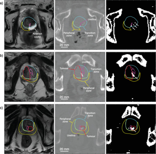
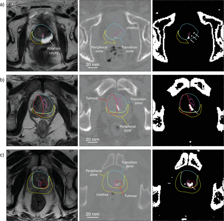
Methods: Patients who underwent both Gallium-68 PSMA PET/CT and MRI of the prostate over the course of a year were selected for analysis. The CT images with visible calcifications within the prostate were included and calcifications automatically isolated using a threshold of 130 HU. The corresponding multiparametric MRI was assessed and the peripheral zone, transition zone, MRI-visible tumor, and urethra manually contoured. The contoured MRI and CT images were registered using rigid registration, and calcifications mapped automatically to the MRI contours.
Results: A total of 85 men (age range 50-88, mean 69 years, standard deviation 7.2 years) were assessed. The mean serum Prostate Specific Antigen PSA was 16.7, range 0.12 to 94.4. Most patients had intermediate-risk disease (68%; Gleason grade group 2 and 3), 26% had high-risk disease (Gleason grade group 4 and 5), and 6% had low-risk disease (Gleason grade group 1). Forty-six patients out of 85 (54%) had intraprostatic calcification. Calcification occurred more in transition zone than the peripheral zone (65% vs. 35%). The mean density of the calcification was 227 HU (min 133, max 1,966 HU). In 12 patients, the calcification was within an MRI-visible tumor, in 24 patients, there were calcifications within a 9 mm distance of the tumor border, and in 9 patients, there were calcifications located between the urethra and tumor.
Conclusions: Calcifications are common in patients with prostate cancer. Their density and location may make them a significant consideration when planning treatment or retreatment with some types of minimally invasive therapy.
Keywords: Calcification; HIFU; PSMA PET; Prostate MRI; Prostate cancer; Radiotherapy.
Copyright © 2021 The Authors. Published by Elsevier Inc. All rights reserved.
Figures
References
-
- Geramoutsos I, Gyftopoulos K, Perimenis P, Thanou V, Liagka D, Siamblis D. Clinical correlation of prostatic lithiasis with chronic pelvic pain syndromes in young adults. Eur Urol. 2004;45(3):333–338. - PubMed
-
- Shoskes DA, Lee CT, Murphy D, Kefer J, Wood HM. Incidence and significance of prostatic stones in men with chronic prostatitis/chronic pelvic pain syndrome. Urology. 2007;70(2):235–238. - PubMed
-
- Fekete CAC, Plamondon M, Martin AG, Vigneault E, Verhaegen F, Beaulieu L. Calcifications in low-dose rate prostate seed brachytherapy treatment: post-planning dosimetry and predictive factors. Radiother Oncol. 2015;114(3):339–344. - PubMed
-
- Georgiou P, Jaros J, Payne H, Allen C, Shah T, Ahmed H. Beam distortion due to gold fiducial markers during salvage high-intensity focused ultrasound in the prostate. Med Phys. 2017;44(2):679–693. - PubMed
Publication types
MeSH terms
Grants and funding
LinkOut - more resources
Full Text Sources
Other Literature Sources
Medical
Research Materials
Miscellaneous
