

Mechanical properties of the different rotator cuff tendons in the rat are similarly and adversely affected by age
- PMID: 33486263
- PMCID: PMC7920936
- DOI: 10.1016/j.jbiomech.2021.110249
Mechanical properties of the different rotator cuff tendons in the rat are similarly and adversely affected by age
Abstract
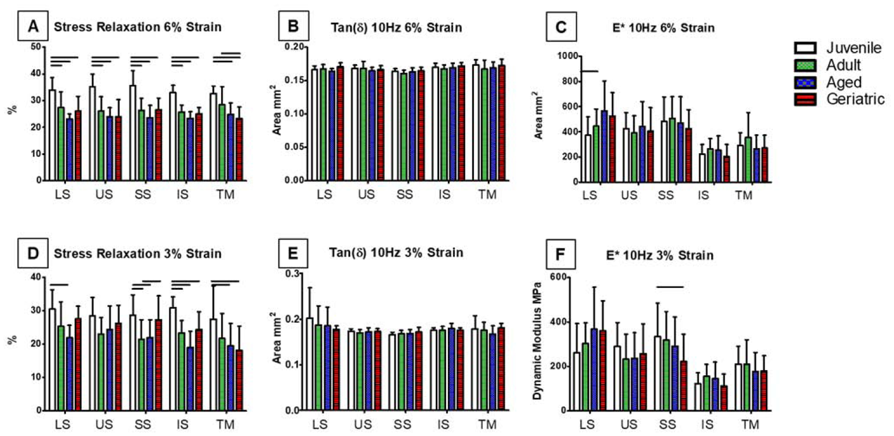
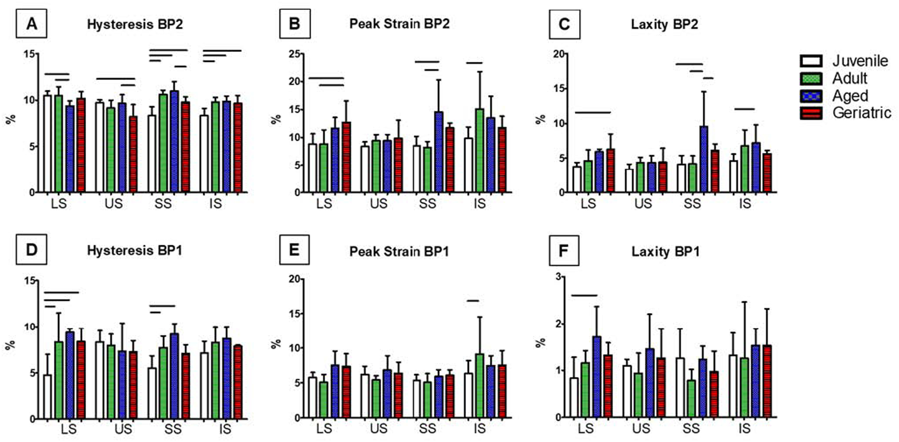
Rotator cuff tendon tears and tendinopathies are common injuries affecting a large portion of the population and can result in pain and joint dysfunction. Incidence of rotator cuff tears significantly increases with advancing age, and up to 90% of these tears involve the supraspinatus. Previous literature has shown that aging can lead to inferior mechanics, altered composition, and changes in structural properties of the supraspinatus. However, there is little known about changes in supraspinatus mechanical properties in context of other rotator cuff tendons. Alterations in tendon mechanical properties may indicate damage and an increased risk of rupture, and thus, the purpose of this study was to use a rat model to define age-related alterations in rotator cuff tendon mechanics to determine why the supraspinatus is more susceptible to tears due to aging than the infraspinatus, subscapularis, and teres minor. Fatigue, viscoelastic, and quasi-static properties were evaluated in juvenile, adult, aged, and geriatric rats. Aging ubiquitously and adversely affected all rotator cuff tendons tested, particularly leading to increased stiffness, decreased stress relaxation, and decreased fatigue secant and tangent moduli in geriatric animals, suggesting a common intrinsic mechanism due to aging in all rotator cuff tendons. This study demonstrates that aging has a significant effect on rotator cuff tendon mechanical properties, though the supraspinatus was not preferentially affected. Thus, we are unable to attribute the aging-associated increase in supraspinatus tears to its mechanical response alone.
Keywords: Aging; Fatigue; Quasi-static; Tendon; Tendon biomechanics; Viscoelasticity.
Copyright © 2021 Elsevier Ltd. All rights reserved.
Conflict of interest statement
CONFLICTS OF INTEREST
The authors have no conflicts of interest to report.
Figures

References
-
- Biberthaler P, Wiedemann E, Nerlich A, Kettler M, Mussack T, Deckelmann S, & Mutschler W, 2003. Microcirculation Associated With Degenerative Rotator Cuff Lesions. In Vivo Assessment With Orthogonal Polarization Spectral Imaging During Arthroscopy of the Shoulder. Journal of Bone and Joint Surgery, 85(3), 475–480. - PubMed
-
- Burkhart S, Morgan C, & Kibler W, 2003. The Disabled Throwing Shoulder: Spectrum of Pathology Part III: The SICK Scapula, Scapular Dyskinesis, the Kinetic Chain, and Rehabilitation. Arthroscopy, 19(6), 641–661. - PubMed
-
- Chen N, & Bedi A, 2010. Rotator Cuff Defect: Acute or Chronic? Journal of Hand Surgery, 36(3), 513–515. - PubMed
-
- Chung S, Park H, Kwon J, Choe G, Kim S, & Oh J, 2016. Effect of Hypercholesterolemia. American Journal of Sports Medicine, 44(5), 1153–1164. - PubMed
Publication types
MeSH terms
Grants and funding
LinkOut - more resources
Full Text Sources
Other Literature Sources
Medical
