. 2020;209(4-6):155-164.
doi: 10.1159/000513040. Epub 2021 Jan 22.

Evidence of SARS-CoV2 Entry Protein ACE2 in the Human Nose and Olfactory Bulb

Affiliations

Evidence of SARS-CoV2 Entry Protein ACE2 in the Human Nose and Olfactory Bulb

Moritz Klingenstein et al. Cells Tissues Organs. 2020.

Abstract

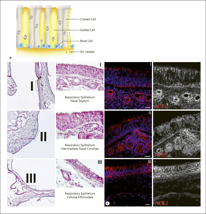
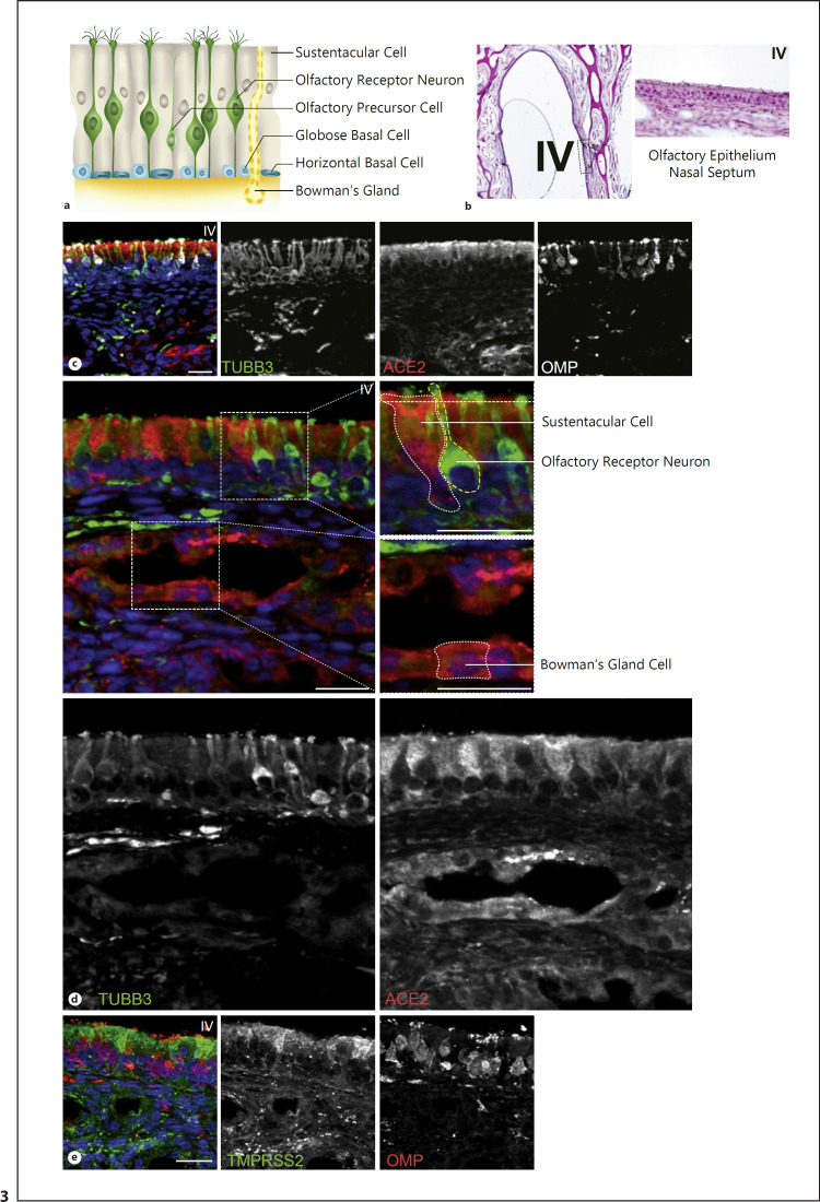
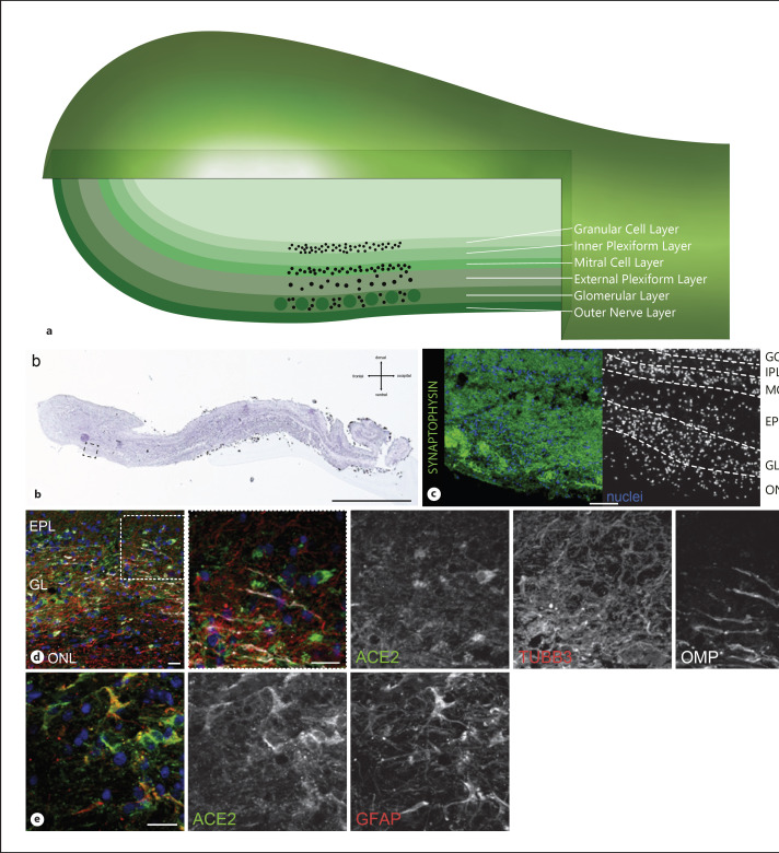
Usually, pandemic COVID-19 disease, caused by SARS-CoV2, presents with mild respiratory symptoms such as fever, cough, but frequently also with anosmia and neurological symptoms. Virus-cell fusion is mediated by angiotensin-converting enzyme 2 (ACE2) and transmembrane serine protease 2 (TMPRSS2) with their organ expression pattern determining viral tropism. Clinical presentation suggests rapid viral dissemination to the central nervous system leading frequently to severe symptoms including viral meningitis. Here, we provide a comprehensive expression landscape of ACE2 and TMPRSS2 proteins across human postmortem nasal and olfactory tissue. Sagittal sections through the human nose complemented with immunolabelling of respective cell types represent different anatomically defined regions including olfactory epithelium, respiratory epithelium of the nasal conchae and the paranasal sinuses along with the hardly accessible human olfactory bulb. ACE2 can be detected in the olfactory epithelium as well as in the respiratory epithelium of the nasal septum, the nasal conchae, and the paranasal sinuses. ACE2 is located in the sustentacular cells and in the glandular cells in the olfactory epithelium as well as in the basal cells, glandular cells, and epithelial cells of the respiratory epithelium. Intriguingly, ACE2 is not expressed in mature or immature olfactory receptor neurons and basal cells in the olfactory epithelium. Similarly, ACE2 is not localized in the olfactory receptor neurons albeit the olfactory bulb is positive. Vice versa, TMPRSS2 can also be detected in the sustentacular cells and the glandular cells of the olfactory epithelium. Our findings provide the basic anatomical evidence for the expression of ACE2 and TMPRSS2 in the human nose, olfactory epithelium, and olfactory bulb. Thus, they are substantial for future studies that aim to elucidate the symptom of SARS-CoV2 induced anosmia via the olfactory pathway.

Keywords: ACE2; Human; Olfactory bulb; Olfactory epithelium; SARS-CoV2.


Conflict of interest statement

The authors have no conflicts of interest to declare.

Figures

Fig. 1
Fig. 1
a Schematic illustration of a human head. Frontal section highlights the nose specimen (a). Sagittal view of the olfactory bulb section (b). Olfactory epithelium and olfactory bulb, both marked in green, are located at the upper nasal cavity and above the cribriform plate. b Schematic representation of the nose specimen from the frontal section through the head (a). The olfactory epithelium (OE) and the olfactory bulb (OB) are shown in green. Section plane for better comprehensibility of the olfactory bulb (b). The dashed boxes show the section of the respiratory epithelium of the nasal septum (NS) (l), the intermediate nasal conchae (INC) (II), the cellulae ethmoidales (CE) (III) and the olfactory epithelium (OE) (IV). CP, cribriform plate; SNC, superior nasal conchae. c. Hematoxylin eosin staining of the right half of the nose specimen. Dashed boxes show the same areas found in the schematic picture in b. d Immunofluorescence staining of the right half of human nose specimen. TUBB3 is shown in green, ACE2 in red, nuclei in blue. Dashed boxes show the same areas found in the schematic picture in b. More details for area I, II, III of the respiratory epithelium can be found in Fig. 2. More details for area IV olfactory epithelium can be found in Fig. 3. Scale bar, 2.5 mm.
Fig. 2
Fig. 2
a Schematic illustration of the pseudostratified respiratory epithelium with ciliated cells, goblet cells, basal cells, and nasal submucosal glands. b Hematoxylin eosin stainings of the respiratory epithelium in the area of the nasal septum (l), the intermediate nasal conchae (II), and the cellulae ethmoidales (III). c Immunofluorescent stainings of the respiratory epithelium verified by absent TUBB3 expression in the epithelium from dashed boxes I, II and III. TUBB3 is a highly specific marker for mature and immature ORN. ACE2 (red) positive basal cells of the respiratory epithelium of the nasal septum as well as positive apical staining of respiratory epithelial cells and nasal submucosal glands (l). ACE2 (red) protein expression is located in the ciliated epithelium cells and the basal cells of the respiratory epithelium of intermediate nasal conchae as well as in the underlying nasal submucosal glands (II). ACE2 (red) expression in the cellulae ethmoidales can be found in epithelial cells and basal cells (III). Nuclei are shown in blue. Scale bar, 20 μm.
Fig. 3
Fig. 3
a Schematic structure of human olfactory epithelium. The mature olfactory receptor neurons are highlighted in green and are surrounded by sustentacular cells. The sensory neurons arise via an olfactory precursor step from basal cells. There are 2 types of basal cells: horizontal and globose basal cells. Bowman glands are secretory glands that are found exclusively in the olfactory epithelium. b Hematoxylin eosin staining of the olfactory epithelium in the area of the nasal septum (IV). c Immunofluorescent staining of the olfactory epithelium from dashed box (IV). Merged picture shows ACE2 in red, OMP, a marker for mature ORN, in white and TUBB3, a marker for mature and immature ORN, in green. Positive ACE2 staining is exclusively located to the region of the sustentacular cells, no appearance in basal cells or co-localization with OMP or TUBB3. d Enlarged picture from the olfactory epithelium and mucosa showing ACE2 positive sustentacular cells and positive Bowman glands. Upper dashed box shows an enlarged picture of ACE2 positive sustentacular cell, marked with dashed white lines, but negative for TUBB3. TUBB3 positive olfactory sensory neuron marked with dashed yellow lines. Lower dashed box shows ACE2 positive staining in Bowman glands in the olfactory submucosa. One ACE2 positive Bowman gland cell is marked with a dashed white line. e Immunofluorescent picture with TMPRSS2 (green) and OMP (red) expression in the olfactory epithelium (IV). TMPRSS2 is mainly located in the sustentacular cells and shows a minor expression in Bowman glands, but no expression in basal cell or OMP-positive mature ORN. Nuclei are shown in blue. Scale bar, 20 μm.
Fig. 4
Fig. 4
a Schematic illustration of the human olfactory bulb with its layers: Under most is the outer nerve layer with the axons of the olfactory receptor neurons followed by the glomerular layer with olfactory glomerula connecting the axons with interneurons. Synaptic processing between the glomerular layer and the mitral cell layer occurs in the external plexiform cell layer. In the mitral cell layer, the mitral cells are located. They form synapses in the inner plexiform layer with the granular cells of the granular cell layer. b Hematoxylin eosin staining of a sagittal section of the human olfactory bulb (b). Dashed box shows an enlarged picture in (c). Scale bar, 2.5 mm. c Immunofluorescence staining of human olfactory bulb. Synaptophysin is shown in green. The specific layers of the olfactory bulb are marked. ONL, outer nerve layer; GL, glomerular layer; EPL, external plexiform layer; MCL, mitral cell layer; IPL, internal plexiform layer; GCL, glomerular cell layer. d Overview picture with immunofluorescence stainings for ACE2 (green), TUBB3 (red), and OMP (white) illustrating the different layers of the olfactory bulb. The white dashed box indicates the magnified area illustrated in the next image, showing representative ACE2 positive cells, not co-localizing with TUBB3 or OMP. High protein expression of TUBB3 and OMP can be found in the outer nerve layer, whereas ACE2 is mainly located in the glomerular layer. e Picture of the olfactory bulb showing ACE2 (green) and glial marker GFAP (red) positive cells. Some GFAP positive glia cells co-localize with ACE2. Nuclei are shown in blue. Scale bar, 20 μm.

Similar articles

Cited by

References

    1. Au WW, Treloar HB, Greer CA. Sublaminar organization of the mouse olfactory bulb nerve layer. J Comp Neurol. 2002;446((1)):68–80. - PubMed
    1. Bilinska K, Jakubowska P, Von Bartheld CS, Butowt R. Expression of the SARS-CoV-2 Entry Proteins, ACE2 and TMPRSS2, in Cells of the Olfactory Epithelium: Identification of Cell Types and Trends with Age. ACS Chem Neurosci. 2020;11((11)):1555–62. - PMC - PubMed
    1. Brann DH, Tsukahara T, Weinreb C, Lipovsek M, Van den Berge K, Gong B, et al. Non-neuronal expression of SARS-CoV-2 entry genes in the olfactory system suggests mechanisms underlying COVID-19-associated anosmia. Sci Adv. 2020;6((31)):eabc5801. - PMC - PubMed
    1. Caggiano M, Kauer JS, Hunter DD. Globose basal cells are neuronal progenitors in the olfactory epithelium: a lineage analysis using a replication-incompetent retrovirus. Neuron. 1994;13((2)):339–52. - PubMed
    1. Chen M, Shen W, Rowan NR, Kulaga H, Hillel A, Ramanathan M, et al. Elevated ACE2 expression in the olfactory neuroepithelium: implications for anosmia and upper respiratory SARS-CoV-2 entry and replication. Eur Respir J. 2020;56:2001948. - PMC - PubMed