

doi: 10.1007/s12104-020-09996-x.
Epub 2021 Jan 24.
NMR assignments of the macro domain from severe acute respiratory syndrome coronavirus 2 (SARS-CoV-2)
Affiliations
- PMID: 33486617
- PMCID: PMC7826497
- DOI: 10.1007/s12104-020-09996-x
Item in Clipboard
NMR assignments of the macro domain from severe acute respiratory syndrome coronavirus 2 (SARS-CoV-2)
Biomol NMR Assign.
2021 Apr.
Abstract
SARS-CoV-2 is a novel pathogen causing pneumonia named COVID-19 and leading to a severe pandemic since the end of 2019. The genome of SARS-CoV-2 contains a macro domain that may play an important role in regulating ADP-ribosylation in host cells and initiating viral replication. Here, we report the 1H, 13C, and 15N resonance assignments of the SARS-CoV-2 macro domain. This work provides the ground for further structural deciphering and biophysical investigation in protein function and antiviral agent design.
Keywords: COVID-19; Macro domain; SARS-CoV-2; Viral protein.
Figures
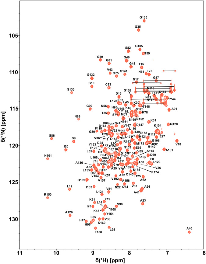
1H-15N HSQC spectrum of SARS-CoV-2 macro domain recorded at 600 MHz with a cryogenic-probe with phosphate buffer pH 6.0 at 298K. Backbone amide 1H and 15N cross-peaks are presented. The horizontal lines connect pairs of the side-chain protons from amino acids Asn and Gln

1H-13C HSQC methyl correlation spectrum of SARS-CoV-2 macro domain recorded at 600 MHz with a cryogenic-probe with phosphate buffer pH 6.0 at 298K. The assigned methyl cross peaks are labeled
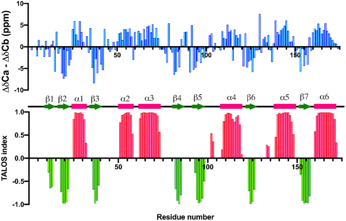
The secondary structure of SARS-CoV-2 macro domain is predicted by CαCβ chemical shift difference, and TALOS+. Upper panel is the parameter ∆δCα − ∆δCβ shows the deviation of Cα and Cβ experimental values from the corresponding random coil values. Positive and negative values suggest α-helix and β-strand structure, respectively. Lower panel is TALOS + index showing the prediction of secondary structure distribution based on backbone N, H, Cα, Hα, C, and side-chain Cβ chemical shift values. Negative and positive values suggest α-helix (in pink) and β-strand (in green) structure, respectively. Chemical shift analysis resulting in secondary structure elements of the macro domain is represented
References
Publication types
MeSH terms
Substances
Grants and funding
LinkOut - more resources
Full Text Sources
Other Literature Sources
Miscellaneous
