Review
. 2021 Mar 15;376(1820):20190758.
doi: 10.1098/rstb.2019.0758. Epub 2021 Jan 25.

Origins of eukaryotic excitability

Affiliations
Review

Origins of eukaryotic excitability

Kirsty Y Wan et al. Philos Trans R Soc Lond B Biol Sci. .

Abstract

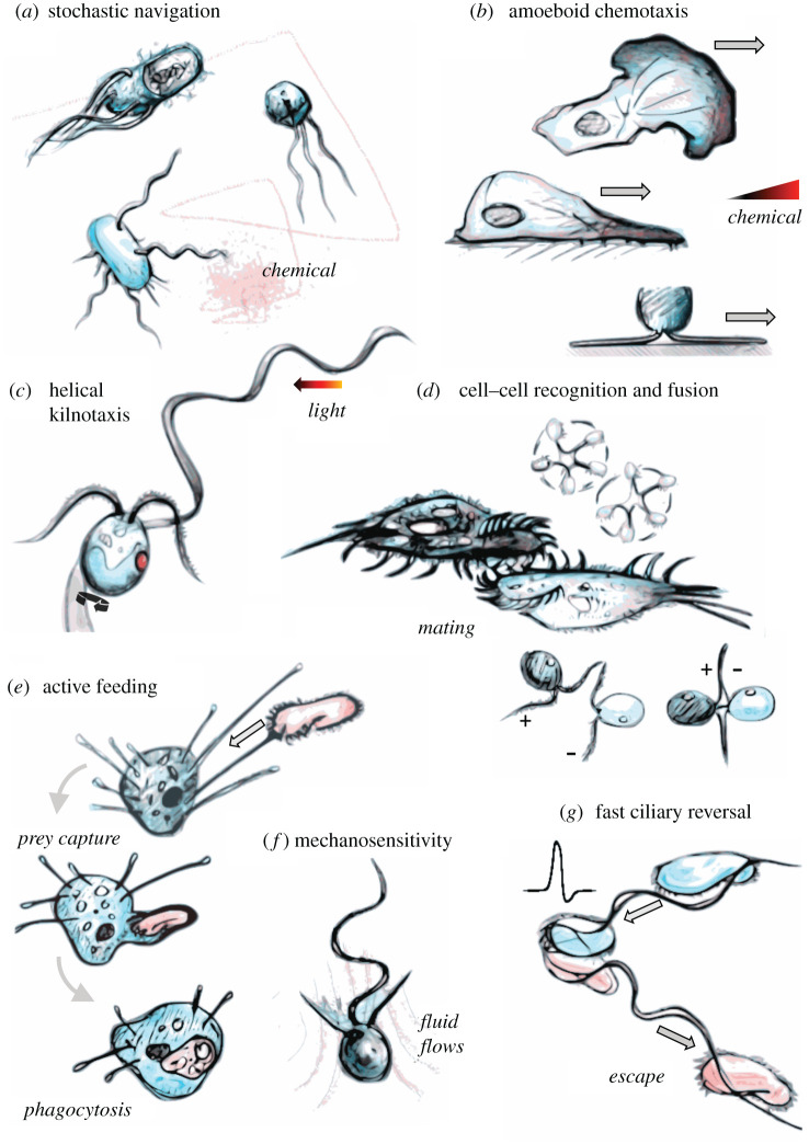
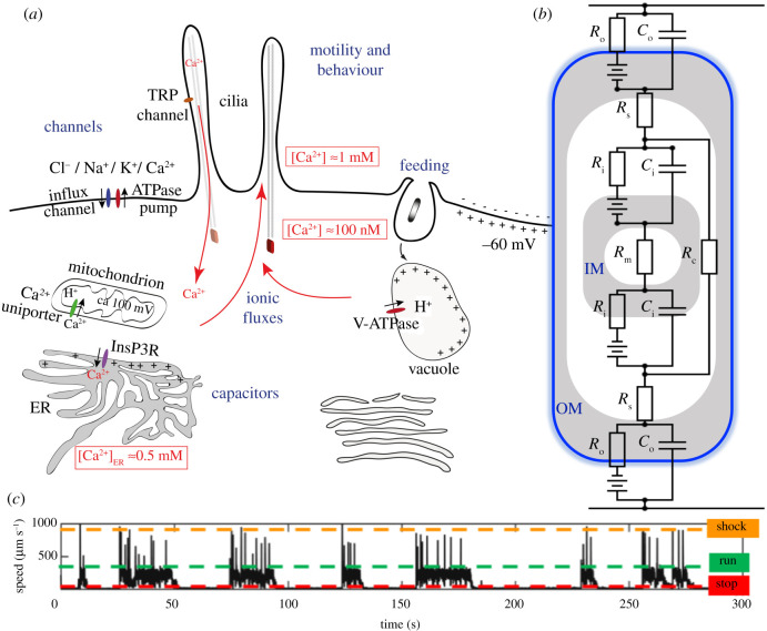
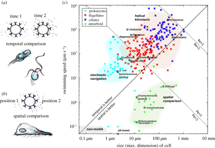
All living cells interact dynamically with a constantly changing world. Eukaryotes, in particular, evolved radically new ways to sense and react to their environment. These advances enabled new and more complex forms of cellular behaviour in eukaryotes, including directional movement, active feeding, mating, and responses to predation. But what are the key events and innovations during eukaryogenesis that made all of this possible? Here we describe the ancestral repertoire of eukaryotic excitability and discuss five major cellular innovations that enabled its evolutionary origin. The innovations include a vastly expanded repertoire of ion channels, the emergence of cilia and pseudopodia, endomembranes as intracellular capacitors, a flexible plasma membrane and the relocation of chemiosmotic ATP synthesis to mitochondria, which liberated the plasma membrane for more complex electrical signalling involved in sensing and reacting. We conjecture that together with an increase in cell size, these new forms of excitability greatly amplified the degrees of freedom associated with cellular responses, allowing eukaryotes to vastly outperform prokaryotes in terms of both speed and accuracy. This comprehensive new perspective on the evolution of excitability enriches our view of eukaryogenesis and emphasizes behaviour and sensing as major contributors to the success of eukaryotes. This article is part of the theme issue 'Basal cognition: conceptual tools and the view from the single cell'.

Keywords: cilia; eukaryogenesis; excitability; membranes; motility; protists.


Figures

Figure 1.
Figure 1.
The many forms of cellular excitability. (a) Stochastic or non-oriented navigation strategies (e.g. prokaryotic chemotaxis), (b) sensing by spatial comparison (e.g. amoeboid chemotaxis), (c) sensing by helical klinotaxis (e.g. flagellate phototaxis), (d) cell–cell recognition as a prelude to fusion (e.g. ciliate conjugation and gametic fusion in Chlamydomonas), (e) active feeding by selective engulfment of prey organisms, (f) mechanosensitivity and flow interactions, and (g) ultrafast escape responses and reversal of ciliary beating by action potentials. (Online version in colour.)
Figure 2.
Figure 2.
Eukaryotic membranes control dynamic intracellular signalling. (a) Massive diversification of lipids and ion channels creates compartments and circuits within a flexible and excitable endomembrane system. (b) The mitochondrion has a distinctive two-layer topology with unique capacitative properties (IM, inner membrane; OM, outer membrane; Ci, Co are IM, OM capacitances; Ri, Ro and Rm are IM, OM and matrix resistances; Rs is the resistance of the inner membrane space; Rc is a ‘shunt’ resistance [264] arising from cristae extending across the intermembrane space). (c) Excitable signalling in cilia manifests as fast transitions or escape responses, as observed here in an octoflagellate protist [228] which switches between three distinct behavioural states (run, stop and shock).
Figure 3.
Figure 3.
Eukaryotes unlocked new biophysical regimes. (a) Temporal sensing strategies typically involve comparing the signal at two slightly different times—this can be stochastic and rotational diffusion limited (as in many prokaryotes), or involve self-steering (as in many ciliates and flagellates). (b) Spatial sensing strategies involve comparing the signal at two different positions at the same time (as in most amoeboid cells). (c) These distinctions create a prokaryote–eukaryote divide with respect to behaviour, as visualized here in a phase space of organism size versus speed (log–log scale). Three phase boundaries partition this space (see also table 2), namely, the Reynolds number (Re), Péclet number (Pe) and the relative advantage of temporal versus spatial sensing (plotted here for an integration time τ = 1 s). Data points are collated from the literature, where some species of particular interest are highlighted. (In particular see: amoeba [326], flagellates and ciliates [327] and marine bacteria [328]. The full dataset is available as supplementary material.)

References

    1. Izhikevich EM. 2010. Dynamical systems in neuroscience: the geometry of excitability and bursting. Cambridge, MA: MIT Press.
    1. Gadsby DC. 2009. Ion channels versus ion pumps: the principal difference, in principle. Nat. Rev. Mol. Cell Biol. 10, 344-352. (10.1038/nrm2668) - DOI - PMC - PubMed
    1. Hodgkin AL, Huxley AF. 1952. A quantitative description of membrane current and its application to conduction and excitation in nerve. J. Physiol. 117, 500-544. (10.1113/jphysiol.1952.sp004764) - DOI - PMC - PubMed
    1. Poincaré H. 1902. La science et l'hypothese [Science and hypothesis], ch. IX. [In French.] Paris, France: Flammarion.
    1. Dusenbery DB. 1992. Sensory ecology: how organisms acquire and respond to information. New York, NY: W. H. Freeman & Company.

Publication types

MeSH terms

LinkOut - more resources