. 2020 Nov-Dec;65(6):452-460.
doi: 10.4103/ijd.IJD_258_20.

Immune Responses in Post Kala-azar Dermal Leishmaniasis

Affiliations

Immune Responses in Post Kala-azar Dermal Leishmaniasis

Mitali Chatterjee et al. Indian J Dermatol. 2020 Nov-Dec.

Abstract

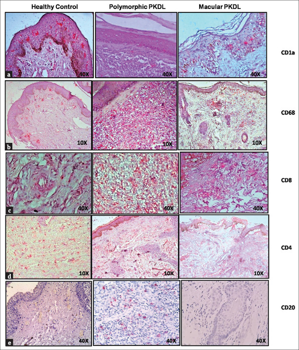
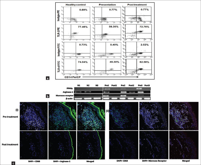
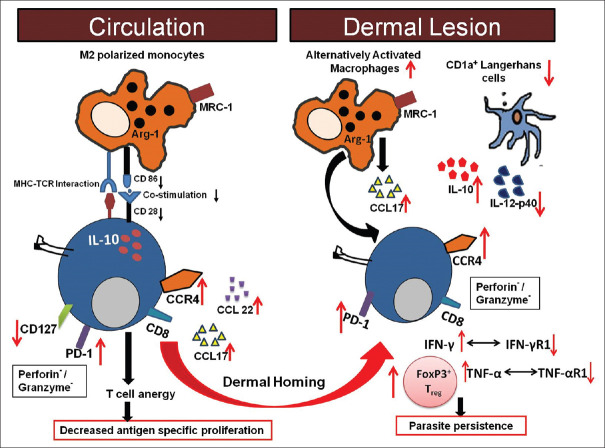
Kala-azar, commonly known as visceral leishmaniasis (VL), is a neglected tropical disease that has been targeted in South Asia for elimination by 2020. Presently, the Kala-azar Elimination Programme is aimed at identifying new low-endemic foci by active case detection, consolidating vector control measures, and decreasing potential reservoirs, of which Post Kala-azar Dermal Leishmaniasis (PKDL) is considered as the most important. PKDL is a skin condition that occurs after apparently successful treatment of VL and is characterized by hypopigmented patches (macular) or a mixture of papules, nodules, and/or macules (polymorphic). To achieve this goal of elimination, it is important to delineate the pathophysiology so that informed decisions can be made regarding the most appropriate and cost-effective approach. We reviewed the literature with regard to PKDL in Asia and Africa and interpreted the findings in establishing a potential correlation between the immune responses and pathophysiology. The overall histopathology indicated the presence of a dense, inflammatory cellular infiltrate, characterized by increased expression of alternatively activated CD68+ macrophages, CD8+ T cells showing features of exhaustion, CD20+ B cells, along with decreased CD1a+ dendritic cells. Accordingly, this review is an update on the overall immunopathology of PKDL, so as to provide a better understanding of host-parasite interactions and the immune responses generated which could translate into availability of markers that can be harnessed for assessment of disease progression and improvement of existing treatment modalities.

Keywords: Immune response; kala-azar; post kala-azar dermal leishmaniasis; visceral leishmaniasis.


Conflict of interest statement

There are no conflicts of interest.

Figures

Figure 1
Figure 1
Distribution of dendritic cells, macrophages, T cells, and B cells in dermal lesions of patients with polymorphic and macular PKDL. Representative immunohistochemical profiles of (a) CD1a, (b) CD68, (c) CD8, (d) CD4 and (e) CD20 from skin biopsies of a healthy control and patient with polymorphic and macular PKDL (×10 and ×40 magnification) (Source: Sengupta R, Mukherjee S, Moulik S, Mitra S, Chaudhuri SJ, Das NK, et al. In-situ immune profile of polymorphic vs. macular Indian Post Kala-azar Dermal Leishmaniasis. Int J Parasitol Drugs Drug Resist 2019;11: 166-76)
Figure 2
Figure 2
(a) Decreased expression of TLR2 and 4 within monocytes. Representative data showing expression of TLR2 and TLR4 within CD14+ monocytes in a healthy control, a patient with PKDL (Pre t/t), and posttreatment (Post t/t). Isotype control staining is also shown. (b) Raised mRNA expression of PPAR-γ, arginase-1 and mannose receptor at the lesional sites in patients with PKDL. Representative mRNA expression profile of PPAR-γ, arginase-1, and mannose receptor in skin samples from healthy controls (N 1–3), patients with PKDL (Pre 1–3), and posttreatment (Post 1–3). The RT-PCR products were quantified by densitometric analysis after normalization with β-actin. (c) Lesional macrophages showed a raised expression of arginase-1 and mannose receptor (i) Expression of arginase-1 (green, Panels 1 and 2) in CD68+ macrophages (white, Panels 1 and 2) at the lesional site of a patient with PKDL (Pre t/t) and posttreatment (Post t/t). Nuclei are shown in blue (DAPI, Panels 1 and 2). Figures were captured in 400X magnification. (ii) Expression of mannose receptor (CD206, white, Panels 1 and 2) in CD68+ macrophages (green, Panels 1 and 2) at the lesional site of a patient with PKDL (Pre t/t) and posttreatment (Post t/t). Nuclei are shown in blue (DAPI, Panels 1 and 2). Figures were captured in 400X magnification. (Source: Mukhopadhyay D, Mukherjee S, Roy S, Dalton JE, Kundu S, Sarkar A, et al. M2 Polarization of monocytes-macrophages is a hallmark of Indian Post Kala-azar Dermal Leishmaniasis. PLoS Negl Trop Dis 2015;9: e0004145)
Figure 3
Figure 3
Cellular interactions between peripheral blood and dermal lesions in patients with PKDL. In patients with PKDL, an increased presence of IL-10 and IL-5 along with immune reinforcement by alternatively activated macrophages (M2) and decreased CD1a+ dendritic cells prevented parasite elimination. Increased CCL17/22 upon interaction with CCR4 led to dermal homing of CD8+ T cells which showed exhaustion as confirmed by the increased presence of PD-1 in peripheral blood and dermal lesions and, benefitted from the milieu generated by the increased presence of IL-4/IL-5 and IL-10

References

    1. Burza S, Croft SL, Boelaert M. Leishmaniasis. Lancet. 2018;392:951–70. - PubMed
    1. Zijlstra EE, Musa AM, Khalil EAG, el-Hassan IM, el-Hassan AM. Post-kala-azar dermal leishmaniasis. Lancet Infect Dis. 2003;3:87–98. - PubMed
    1. Zijlstra EE. The immunology of post-kala-azar dermal leishmaniasis (PKDL) Parasit Vectors. 2016;9:464. - PMC - PubMed
    1. Ganguly S, Das NK, Barbhuiya JN, Chatterjee M. Post-kala-azar dermal leishmaniasis-an overview. Int J Dermatol. 2010;49:921–31. - PubMed
    1. Ramesh V, Kaushal H, Mishra AK, Singh R, Salotra P. Clinico-epidemiological analysis of Post kala-azar dermal leishmaniasis (PKDL) cases in India over last two decades: A hospital based retrospective study. BMC Public Health. 2015;15:1092. - PMC - PubMed