

A 16-year Survey of Clinicopathological Findings, Electron Microscopy, and Classification of Renal Amyloidosis
- PMID: 33487790
- PMCID: PMC7812497
- DOI: 10.30476/ijms.2019.82110.
A 16-year Survey of Clinicopathological Findings, Electron Microscopy, and Classification of Renal Amyloidosis
Abstract
Background: Electron microscopy (EM) is a valuable tool in the diagnosis of renal amyloidosis, particularly in the early stages of the disease. In Iran, studies on EM and the clinical features of renal amyloidosis are scarce. The objective of the present study was to survey EM investigations, pathological classifications, and clinical features of renal amyloidosis.
Methods: This cross-sectional study was performed in Shiraz, Iran, during 2001-2016. Out of 2,770 kidney biopsies, 27 cases with a diagnosis of renal amyloidosis were investigated. EM investigation and six staining procedures for light microscopy (LM) were performed. Two pathological classifications based on glomerular, peritubular, perivascular, and interstitial involvement were made. Finally, the association between these classifications and the clinical features was assessed. Chi-square, Fisher's exact, Independent t test, and Multiple logistic regression analysis were used. P values<0.05 were considered statistically significant.
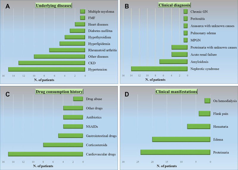
Results: In 51.9% of the cases, the clinical diagnosis was nephrotic syndrome. Proteinuria and edema were the most prevalent clinical manifestations. The role of EM investigation for diagnosis was graded "necessary" or "supportive" in 48.2% of the patients. In the classification based on glomerular classes, variables such as the mean blood pressure (P=0.003), history of hypertension (P=0.02), creatinine >1.5 (P=0.03), and severe tubular atrophy (P=0.03) were significantly higher in class B (advanced amyloid depositions).
Conclusion: EM plays an important role in the diagnosis of renal amyloidosis. EM in conjunction with LM investigation with Congo red staining is recommended, to prevent misdiagnosis of patients with a clinical suspicion of renal amyloidosis. Among different pathological features of renal amyloidosis, the severity of glomerular amyloid depositions had a clear relationship with clinical presentations.
Keywords: Amyloidosis; Electrons; Kidney; Microscopy; Nephrotic syndrome; Proteinuria.
Copyright: © Iranian Journal of Medical Sciences.
Figures


Similar articles
-
[The clinicopathological features of early renal amyloidosis].Zhonghua Bing Li Xue Za Zhi. 2003 Apr;32(2):120-3. Zhonghua Bing Li Xue Za Zhi. 2003. PMID: 12839672 Chinese.
-
Prevalence and origin of amyloid in kidney biopsies.Am J Surg Pathol. 2009 Aug;33(8):1198-205. doi: 10.1097/PAS.0b013e3181abdfa7. Am J Surg Pathol. 2009. PMID: 19561448
-
Analysis of clinical and pathological characteristics of 28 cases with renal amyloidosis.Clin Lab. 2011;57(11-12):947-52. Clin Lab. 2011. PMID: 22239026
-
[Renal disorders associated with monoclonal gammopathies: diagnostic and therapeutic progress].Presse Med. 2012 Mar;41(3 Pt 1):276-89. doi: 10.1016/j.lpm.2011.11.008. Epub 2012 Jan 13. Presse Med. 2012. PMID: 22244725 Review. French.
-
[Clinical features of patients with the pathological diagnosis of amyloidosis].Rev Med Chil. 2005 Jun;133(6):655-61. doi: 10.4067/s0034-98872005000600006. Epub 2005 Jul 22. Rev Med Chil. 2005. PMID: 16075129 Review. Spanish.
Cited by
-
Renal Amyloidosis: A Clinicopathological Study From a Tertiary Care Hospital in Pakistan.Cureus. 2022 Jan 11;14(1):e21122. doi: 10.7759/cureus.21122. eCollection 2022 Jan. Cureus. 2022. PMID: 35165578 Free PMC article.
-
Research trends and hotspots evolution of cardiac amyloidosis: a bibliometric analysis from 2000 to 2022.Eur J Med Res. 2023 Feb 20;28(1):89. doi: 10.1186/s40001-023-01026-5. Eur J Med Res. 2023. PMID: 36805827 Free PMC article. Review.
References
-
- Bustamante JG, Gossman WG. Amyloidosis. StatPearls: StatPearls Publishing; 2019.
-
- Said SM, Sethi S, Valeri AM, Leung N, Cornell LD, Fidler ME, et al. Renal amyloidosis: origin and clinicopathologic correlations of 474 recent cases. Clin J Am Soc Nephrol. 2013;8:1515–23. doi: 10.2215/CJN.10491012. [ PMC Free Article ] - DOI - PMC - PubMed
-
- Gertz MA. Amyloidosis: diagnosis and prognosis. International Journal of Clinical Rheumatology. 2008;3:369. doi: 10.2217/17460816.3.4.369. - DOI
Publication types
MeSH terms
LinkOut - more resources
Full Text Sources
Medical