. 2020 Dec 7:2020:1678780.
doi: 10.1155/2020/1678780. eCollection 2020.

Detection of CD39 and a Highly Glycosylated Isoform of Soluble CD73 in the Plasma of Patients with Cervical Cancer: Correlation with Disease Progression

Affiliations

Detection of CD39 and a Highly Glycosylated Isoform of Soluble CD73 in the Plasma of Patients with Cervical Cancer: Correlation with Disease Progression

Ricardo Muñóz-Godínez et al. Mediators Inflamm. .

Abstract

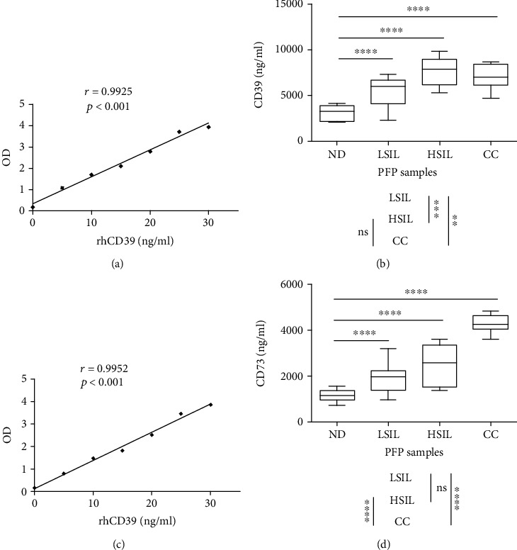
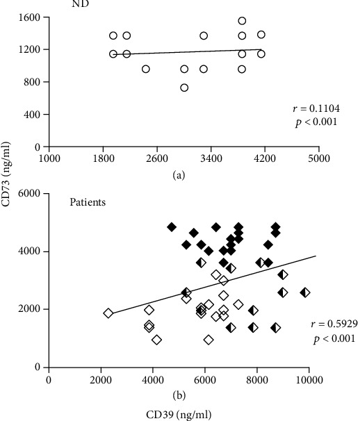
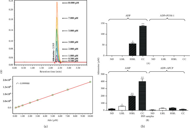
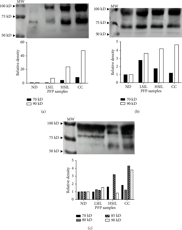
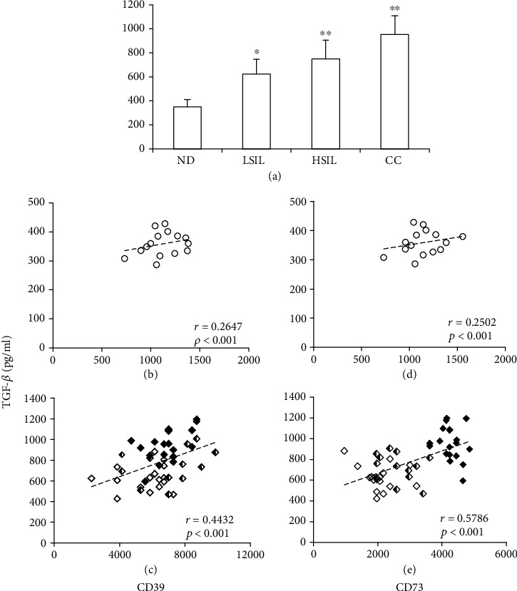
Persistent infection with high-risk human papillomavirus (HR-HPV) is the main factor in the development of cervical cancer (CC). The presence of immunosuppressive factors plays an important role in the development of this type of cancer. To determine whether CD39 and CD73, which participate in the production of immunosuppressive adenosine (Ado), are involved in the progression of CC, we compared the concentrations and hydrolytic activity of these ectonucleotidases in platelet-free plasma (PFP) samples between patients with low-grade squamous intraepithelial lesions (LSILs) (n = 18), high-grade squamous intraepithelial lesions (HSILs) (n = 12), and CC (n = 19) and normal donors (NDs) (n = 15). The concentrations of CD39 and CD73 in PFP increased with disease progression (r = 0.5929, p < 0.001). The PFP of patients with HSILs or CC showed the highest concentrations of CD39 (2.3 and 2.2 times that of the NDs, respectively) and CD73 (1.7 and 2.68 times that of the NDs, respectively), which were associated with a high capacity to generate Ado from the hydrolysis of adenosine diphosphate (ADP) and adenosine monophosphate (AMP). The addition of POM-1 and APCP, specific inhibitors of CD39 and CD73, respectively, inhibited the ADPase and AMPase activity of PFP by more than 90%. A high level of the 90 kD isoform of CD73 was detected in the PFP of patients with HSILs or CC. Digestion with endoglycosidase H and N-glycanase generated CD73 with weights of approximately 90 kD, 85 kD, 80 kD, and 70 kD. In addition, the levels of transforming grow factor-β (TGF-β) in the PFPs of patients with LSIL, HSIL and CC positively correlated with those of CD39 (r = 0.4432, p < 0.001) and CD73 (r = 0.5786, p < 0.001). These results suggest that persistent infection by HR-HPV and the concomitant production of TGF-β promote the expression of CD39 and CD73 to favor CC progression through Ado generation.


Conflict of interest statement

The authors have no conflicts of interest to declare.

Figures

Figure 1
Figure 1
CD39 and CD73 concentrations in PFP samples from NDs and patients with LSILs, HSILs, or CC. The type curves for soluble CD39 and CD73 were established by ELISA using different concentrations (5-30 ng/ml) of human recombinant proteins CD39 (rhCD39) (a) and CD73 (rhCD73) (c), whose correlations with absorbance (optical density) were r = 0.9925 (p < 0.001) and r = 0.9952 (p < 0.001), respectively. For the detection of CD39 and CD73, the PFP samples of the NDs and women with LSILs, HSILs, or CC were diluted with PBS at ratios of 1 : 40,000 and 1 : 25,000, respectively. The data are representative of three independent experiments. The means ± SEMs of the concentrations of soluble CD39 (b) and CD73 (d) detected in the PFP of the NDs and patients with LSILs, HSILs, or CC are shown. Significant differences are indicated by ∗ (p < 0.05), ∗∗ (p < 0.001), and ∗∗∗ (p < 0.0001). ns: not significant.
Figure 2
Figure 2
Correlations between the concentrations of CD39 and CD73 in the PFP of NDs and in the PFP of patients (LSILs, HSILs, and CC together). The correlations (r) between the concentrations of CD39 and CD73 in the ND PFP samples (open circles) are shown (r = 0.1104, p < 0.001) (a), as well as the correlations for patients with LSILs (white diamonds), HSILs (black and white diamonds), and CC (black diamonds) (b) (r = 0.5929, p < 0.001).
Figure 3
Figure 3
Catalytic activity of CD39 and CD73 in PFP of NDs and patients with LSILs, HSILs, or CC. Aliquots of 5 μl of PFP from NDs (black bars), patients with LSILs (white bars), patients with HSILs (diagonal lines), and patients with CC (horizontal lines) were incubated in the presence of 5 mM ADP or AMP (total volume 100 μl) and in the presence or absence of POM-1 or APCP, specific inhibitors of CD39 and CD73, respectively. (a) The Ado produced by hydrolysis of the nucleotides was quantified after 72 h by UPLC using standard concentrations of synthetic Ado (upper). A representative linear relationship between the Ado concentration and the optical density is shown (lower). (b) The amount of Ado produced during the incubation of PFP with ADP (upper) or AMP (lower) and in the presence or absence of POM-1 or APCP is shown. Differences in Ado concentrations were analyzed by two-way ANOVA. p < 0.01, ∗∗p < 0.0001. The data represent three independent experiments.
Figure 4
Figure 4
Detection of CD73 in the PFP of NDs and patients with LSILs, HSILs, or CC. Samples of 3 μl of PFP (a) or 20 ng of CD73 contained in the PFP (b) of the ND, LSIL, HSIL, and CC groups were analyzed by Western blot using the anti-CD73 antibody. The densities of the 70 kD (black bars) and 90 kD (white bars) bands detected in the samples of patients with LSILs, HSILs, or CC relative to those of NDs (set to 1) are shown. (c) Twenty-nanogram samples of CD73 contained in PFP were subjected to deglycosylation using the enzymes endoglycosidase H and N-glycanase. The densities of the 70 kD (black bars), 80 kD (bars with horizontal lines), 85 kD (bars with diagonals), and 90 kD (white bars) bands detected in the LSIL, HSIL, and CC samples relative to the ND samples (set to 1) are shown. MW: molecular weight. A representative test of three independent tests is shown.
Figure 5
Figure 5
TGF-β concentrations in PFP samples from NDs and patients with LSILs, HSILs, or CC and their correlations with the concentrations of CD39 and CD73. (a) The TGF-β1 content is shown in PFP samples taken from NDs and patients with LSILs, HSILs, or CC. The data are representative of three independent experiments, and the means ± SEMs are shown. p < 0.05, ∗∗p < 0.001 for the concentration of TGF-β1 in a patient group relative to the ND group. The correlations (r) between the levels of TGF-β1 and CD39 or CD73 in the PFP of NDs were r = 0.2647 (p < 0.001) (b) and r = 0.2502 (p < 0.001) (d), respectively. For patients with LSILs, HSILs, or CC, the correlations were r = 0.4432 (p < 0.001) (c) and r = 0.5786 (p < 0.001) (e), respectively. The coordinates of the TGF-β1 concentration with the CD39 or CD73 concentration are indicated by white circles in the ND group, white diamonds in the LSIL group, white and black diamonds in the HSIL group, and black diamonds in the CC group.

References

    1. Bray F., Ferlay J., Soerjomataram I., Siegel R. L., Torre L. A., Jemal A. Global cancer statistics 2018: GLOBOCAN estimates of incidence and mortality worldwide for 36 cancers in 185 countries. CA: A Cancer Journal of Clinicians. 2018;68(6):394–424. - PubMed
    1. Woodman C., Collins S., Young L. The natural history of cervical HPV infection: unresolved issues. Nature Reviews Cancer. 2007;7(1):11–22. doi: 10.1038/nrc2050. - DOI - PubMed
    1. IARC Working Group. Biological agents. Volume 100 B. A review of human carcinogens. IARC monographs on the evaluation of carcinogenic risks to humans/World Health Organization, International Agency for Research on Cancer. 2012;100:1–441.
    1. Muñoz N., Bosch F. X., de Sanjosé S., et al. Epidemiologic classification of human papillomavirus types associated with cervical cancer. New England Journal of Medicine. 2003;348(6):518–527. doi: 10.1056/NEJMoa021641. - DOI - PubMed
    1. Westrich J., Warren C., Pyeon D. Evasion of host immune defenses by human papillomavirus. Virus Research. 2017;231(2):21–33. doi: 10.1016/j.virusres.2016.11.023. - DOI - PMC - PubMed