

Pathophysiological Processes Underlying the High Prevalence of Deep Vein Thrombosis in Critically Ill COVID-19 Patients
- PMID: 33488398
- PMCID: PMC7820906
- DOI: 10.3389/fphys.2020.608788
Pathophysiological Processes Underlying the High Prevalence of Deep Vein Thrombosis in Critically Ill COVID-19 Patients
Abstract
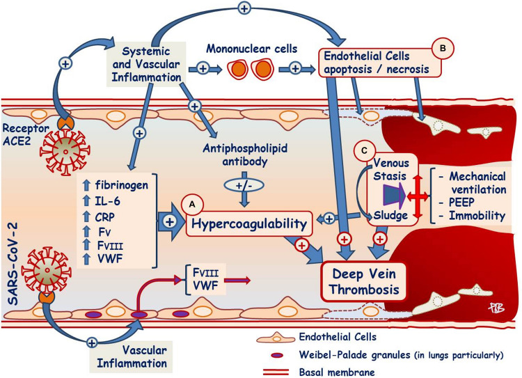
Coronavirus disease 2019 (COVID-19) predisposes to deep vein thrombosis (DVT) and pulmonary embolism (PE) particularly in mechanically ventilated adults with severe pneumonia. The extremely high prevalence of DVT in the COVID-19 patients hospitalized in the intensive care unit (ICU) has been established between 25 and 84% based on studies including systematic duplex ultrasound of the lower limbs when prophylactic anticoagulation was systematically administrated. DVT prevalence has been shown to be markedly higher than in mechanically ventilated influenza patients (6-8%). Unusually high inflammatory and prothrombotic phenotype represents a striking feature of COVID-19 patients, as reflected by markedly elevated reactive protein C, fibrinogen, interleukin 6, von Willebrand factor, and factor VIII. Moreover, in critically ill patients, venous stasis has been associated with the prothrombotic phenotype attributed to COVID-19, which increases the risk of thrombosis. Venous stasis results among others from immobilization under muscular paralysis, mechanical ventilation with high positive end-expiratory pressure, and pulmonary microvascular network injuries or occlusions. Venous return to the heart is subsequently decreased with increase in central and peripheral venous pressures, marked proximal and distal veins dilation, and drops in venous blood flow velocities, leading to a spontaneous contrast "sludge pattern" in veins considered as prothrombotic. Together with endothelial lesions and hypercoagulability status, venous stasis completes the Virchow triad and considerably increases the prevalence of DVT and PE in critically ill COVID-19 patients, therefore raising questions regarding the optimal doses for thromboprophylaxis during ICU stay.
Keywords: COVID-19; D-dimer; deep vein thrombosis; hemostasis disorder; venous stasis.
Copyright © 2021 Voicu, Ketfi, Stépanian, Chousterman, Mohamedi, Siguret, Mebazaa, Mégarbane and Bonnin.
Conflict of interest statement
AM reports personal fees from Orion, Servier, Otsuka, Philips, Sanofi, Adrenomed, Epygon, and Fire 1 and grants and personal fees from 4TEEN4, Abbott, Roche, and Sphingotec. The remaining authors declare that the research was conducted in the absence of any commercial or financial relationships that could be construed as a potential conflict of interest.
Figures
References
-
- Blasi A., von Meijenfeldt F. A., Adelmeijer J., Calvo A., Ibañez C., Perdomo J., et al. (2020). In vitro hypercoagulability and ongoing in vivo activation of coagulation and fibrinolysis in COVID-19 patients on anticoagulation. J. Thromb. Haemost. 18 2646–2653. 10.1111/jth.15043 - DOI - PMC - PubMed
Publication types
LinkOut - more resources
Full Text Sources
Other Literature Sources