

A large Middle Devonian eubrachythoracid 'placoderm' (Arthrodira) jaw from northern Gondwana
- PMID: 33488510
- PMCID: PMC7809001
- DOI: 10.1186/s13358-020-00212-w
A large Middle Devonian eubrachythoracid 'placoderm' (Arthrodira) jaw from northern Gondwana
Abstract
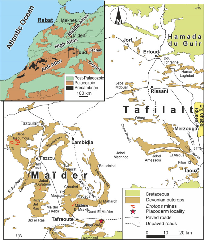
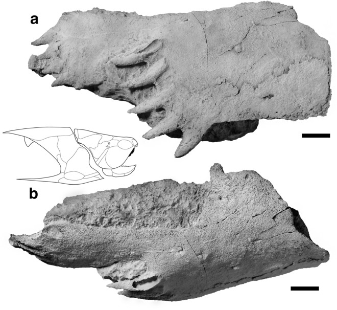
For the understanding of the evolution of jawed vertebrates and jaws and teeth, 'placoderms' are crucial as they exhibit an impressive morphological disparity associated with the early stages of this process. The Devonian of Morocco is famous for its rich occurrences of arthrodire 'placoderms'. While Late Devonian strata are rich in arthrodire remains, they are less common in older strata. Here, we describe a large tooth-bearing jaw element of Leptodontichthys ziregensis gen. et sp. nov., an eubrachythoracid arthrodire from the Middle Devonian of Morocco. This species is based on a large posterior superognathal with a strong dentition. The jawbone displays features considered synapomorphies of Late Devonian eubrachythoracid arthrodires, with one posterior and one lateral row of conical teeth oriented postero-lingually. μCT-images reveal internal structures including pulp cavities and dentinous tissues. The posterior orientation of the teeth and the traces of a putative occlusal contact on the lingual side of the bone imply that these teeth were hardly used for feeding. Similar to Compagopiscis and Plourdosteus, functional teeth were possibly present during an earlier developmental stage and have been worn entirely. The morphological features of the jaw element suggest a close relationship with plourdosteids. Its size implies that the animal was rather large.
Keywords: Arthrodira; Dentition; Food web; Givetian; Maïder basin; Palaeoecology.
© The Author(s) 2021.
Conflict of interest statement
Competing InterestsThe authors declare that they have no competing interests.
Figures


Similar articles
-
A well-preserved 'placoderm' (stem-group Gnathostomata) upper jaw from the Early Devonian of Mongolia clarifies jaw evolution.R Soc Open Sci. 2023 Feb 22;10(2):221452. doi: 10.1098/rsos.221452. eCollection 2023 Feb. R Soc Open Sci. 2023. PMID: 36844806 Free PMC article.
-
Development of teeth and jaws in the earliest jawed vertebrates.Nature. 2012 Nov 29;491(7426):748-51. doi: 10.1038/nature11555. Epub 2012 Oct 17. Nature. 2012. PMID: 23075852
-
Extreme lower jaw elongation in a placoderm reflects high disparity and modularity in early vertebrate evolution.R Soc Open Sci. 2024 Jan 31;11(1):231747. doi: 10.1098/rsos.231747. eCollection 2024 Jan. R Soc Open Sci. 2024. PMID: 38298398 Free PMC article.
-
Origin and evolution of gnathostome dentitions: a question of teeth and pharyngeal denticles in placoderms.Biol Rev Camb Philos Soc. 2005 May;80(2):303-45. doi: 10.1017/s1464793104006682. Biol Rev Camb Philos Soc. 2005. PMID: 15921053 Review.
-
Conserved developmental processes constrain evolution of lungfish dentitions.J Anat. 2001 Jul-Aug;199(Pt 1-2):161-8. doi: 10.1046/j.1469-7580.2001.19910161.x. J Anat. 2001. PMID: 11523818 Free PMC article. Review.
Cited by
-
Diverse stem-chondrichthyan oral structures and evidence for an independently acquired acanthodid dentition.R Soc Open Sci. 2021 Nov 10;8(11):210822. doi: 10.1098/rsos.210822. eCollection 2021 Nov. R Soc Open Sci. 2021. PMID: 34804566 Free PMC article.
-
Reconstruction of feeding behaviour and diet in Devonian ctenacanth chondrichthyans using dental microwear texture and finite element analyses.R Soc Open Sci. 2025 Jan 29;12(1):240936. doi: 10.1098/rsos.240936. eCollection 2025 Jan. R Soc Open Sci. 2025. PMID: 39881788 Free PMC article.
References
-
- Anderson, P. S. L. (2008). Shape variation between arthrodire morphotypes indicates possible feeding niches. Journal of Vertebrate Paleontology,28, 961–969. 10.1671/0272-4634-28.4.961.
-
- Anderson, P. S. L. (2010). Using linkage models to explore skull kinematic diversity and functional convergence in arthrodire placoderms. Journal of Morphology. 10.1002/jmor.10850. - PubMed
-
- Blieck, A., Golshani, F., Goujet, D., Hamdi, A., Janvier, P., Mark-Kurik, E., & Martin, M. (1980). A new vertebrate locality in the Eifelian of the Khush-Yeilagh Formation, Eastern Alborz. Iran. Palaeovertebrata,9, 133–154.
-
- Blieck, A., & Goujet, D. (1991). Les vertébrés du Dévonien inférieur d’Arville et de Nonceveux (Belgique). Annales de la Société géologique du Nord,1, 67–78.
LinkOut - more resources
Full Text Sources
Other Literature Sources