Review
. 2021 Jan 8:11:616020.
doi: 10.3389/fimmu.2020.616020. eCollection 2020.

Antigen-Presenting Cells in Food Tolerance and Allergy

Affiliations
Review

Antigen-Presenting Cells in Food Tolerance and Allergy

Elise G Liu et al. Front Immunol. .

Abstract

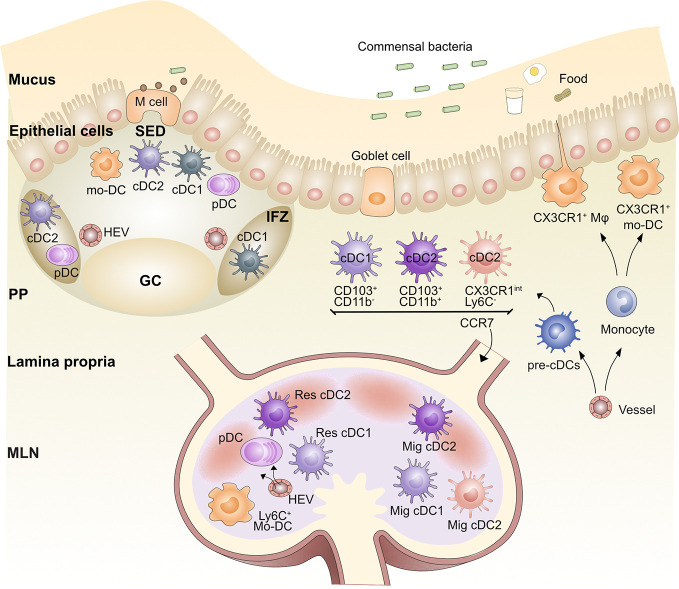
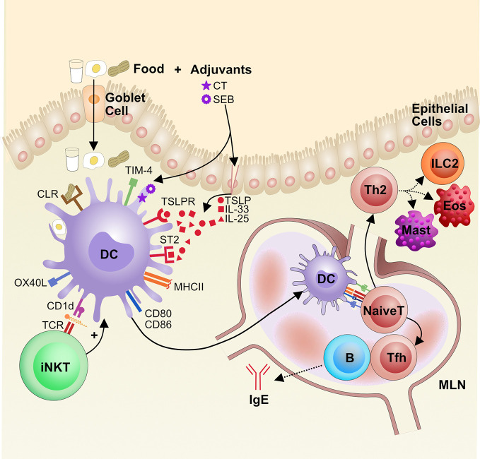
Food allergy now affects 6%-8% of children in the Western world; despite this, we understand little about why certain people become sensitized to food allergens. The dominant form of food allergy is mediated by food-specific immunoglobulin E (IgE) antibodies, which can cause a variety of symptoms, including life-threatening anaphylaxis. A central step in this immune response to food antigens that differentiates tolerance from allergy is the initial priming of T cells by antigen-presenting cells (APCs), primarily different types of dendritic cells (DCs). DCs, along with monocyte and macrophage populations, dictate oral tolerance versus allergy by shaping the T cell and subsequent B cell antibody response. A growing body of literature has shed light on the conditions under which antigen presentation occurs and how different types of T cell responses are induced by different APCs. We will review APC subsets in the gut and discuss mechanisms of APC-induced oral tolerance versus allergy to food identified using mouse models and patient samples.

Keywords: Peyer’s patches; dendritic cells; food allergy; gut; macrophages; mesenteric lymph node; monocytes; oral tolerance.


Conflict of interest statement

The authors declare that the research was conducted in the absence of any commercial or financial relationships that could be construed as a potential conflict of interest.

Figures

Figure 1
Figure 1
Organization of the gut antigen presenting cell network. Blood pre-cDCs populate the lamina propria (LP), Peyer’s Patches (PP), and mesenteric lymph node (MLN) and differentiate into cDC1s and cDC2s. After being activated by antigen, LP cDC1s and cDC2s are able to migrate via afferent lymphatics to the gut-draining MLN via CCR7; these DCs are called migratory DCs (Mig DC). Similarly, cDC2s and possibly cDC1s in the subepithelial dome (SED) of the PPs are able to migrate to the intrafollicular zone (IFZ). Lysozyme+CX3CR1+ monocyte-derived DCs (mo-DC) also populate the SED. Pre-cDCs travel through the blood and seed the MLN and PP, where they differentiate into resident (Res) cDC1 and Res cDC2. Plasmacytoid DCs (pDCs) also populate the LP, PP, and MLN. Blood-derived monocytes differentiate into LP and PP macrophages (Mφ) as well as mo-DCs. Germinal center (GC), Microfold (M) cell, High endothelial venule (HEV).
Figure 2
Figure 2
Mechanisms by which gut-associated dendritic cells contribute to oral tolerance. After food ingestion, goblet cells and intestinal resident macrophages sample luminal food antigens and deliver them to LP CD103+ cDCs (including CD103+CD11b-cDC1s and CD103+CD11b+ cDC2s). Commensal bacterial metabolites, dietary components such as vitamin A, and epithelial cell-derived TGF-β and retinoic acid (RA) imprint tolerogenic properties on cDCs. These cDCs migrate to MLNs through afferent lymphatic vessels and induce naïve CD4+ T cells to differentiate into peripheral regulatory T (pTreg) cells through TGF-β and RA. CD103+ cDCs induce gut homing molecules CCR9 and α4β7 on pTreg cells, which directs them to recirculate to intestinal tissue. Once in the lamina propria, pTreg cells can be further expanded by macrophages (Mφ), possibly via IL-10 production. MLN, mesenteric lymph node; RA, retinoic acid. Conventional dendritic cell (cDC), Innate lymphoid cell type 3 (ILC3).
Figure 3
Figure 3
The role of dendritic cells in the pathogenesis of food allergy. Food antigens are taken up from the gut lumen by goblet cells, which shuttle the antigens across the epithelial layer to the LP, where local dendritic cells (DCs) sample the food antigens. If DCs sense innate immune signals, adjuvants that are either extrinsic or intrinsic to the food antigen, they become activated. Some adjuvants damage the epithelial barrier and trigger the release of alarmins, like TSLP and IL-33, that can activate DCs via their receptors TSLPR and ST2, respectively. Additionally, food glycoproteins, such as from peanut, can bind to C-type lectin receptors (CLRs) and activate DCs. Lipids from foods can be presented on CD1d to iNKT cells that then reciprocally activate DCs via cytokine release. Activation of DCs leads to increased CCR7 for migration to mesenteric lymph nodes (MLN) along with presentation of food antigens on MHCII and increased expression of costimulatory molecules CD80, CD86, OX40L, and TIM-4. Altogether this promotes naïve CD4+ T cell priming and differentiation into Th2 cells and T follicular helper (Tfh) cells, which drive cellular and IgE responses in food allergy, respectively. Eos, eosinophils (Eos), Mast cell (Mast), Innate lymphoid cells type 2 (ILC2).

References

    1. Gupta RS, Springston EE, Warrier MR, Smith B, Kumar R, Pongracic J, et al. The Prevalence, Severity, and Distribution of Childhood Food Allergy in the United States. Pediatrics (2011) 128(1):e9–17. 10.1542/peds.2011-0204 - DOI - PubMed
    1. Sicherer SH. Epidemiology of food allergy. J Allergy Clin Immunol (2011) 127(3):594–602. 10.1016/j.jaci.2010.11.044 - DOI - PubMed
    1. Tang MLK, Mullins RJ. Food allergy: is prevalence increasing? Internal Med J (2017) 47(3):256–61. 10.1111/imj.13362 - DOI - PubMed
    1. Branum, Lukacs SL. Food Allergy Among Children in the United States. Pediatrics (2009) 124(6):1549–55. 10.1542/peds.2009-1210 - DOI - PubMed
    1. Sicherer SH, Sampson HA. 9. Food allergy. J Allergy Clin Immunol (2006) 117(2, Supplement 2):S470–5. 10.1016/j.jaci.2005.05.048 - DOI - PubMed

Publication types