Review
. 2020 Dec 10:2020:8810813.
doi: 10.1155/2020/8810813. eCollection 2020.

Adipose-Derived Stem Cells: Current Applications and Future Directions in the Regeneration of Multiple Tissues

Affiliations
Review

Adipose-Derived Stem Cells: Current Applications and Future Directions in the Regeneration of Multiple Tissues

Jiaxin Zhang et al. Stem Cells Int. .

Abstract

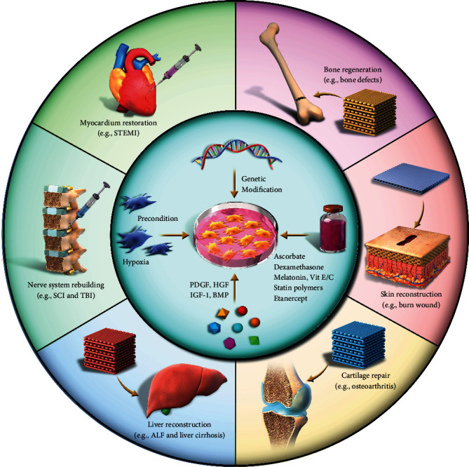
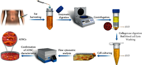
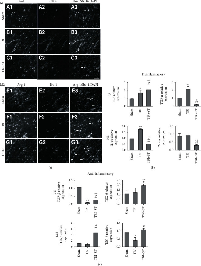
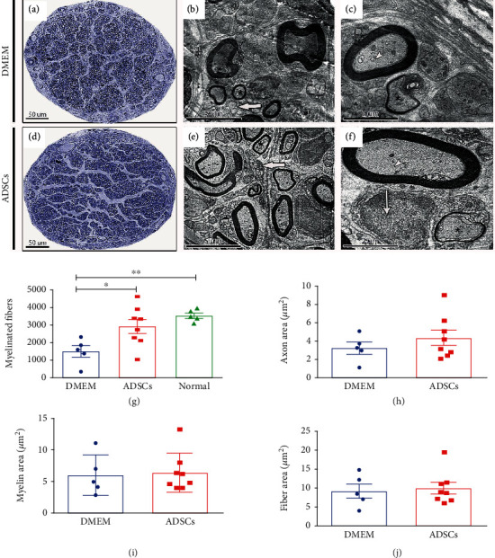
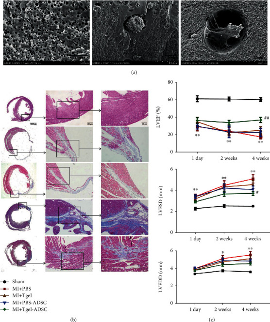
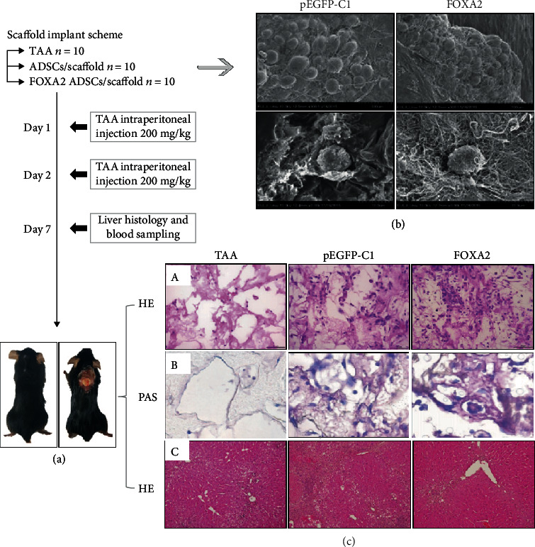
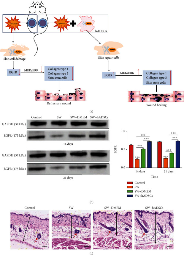
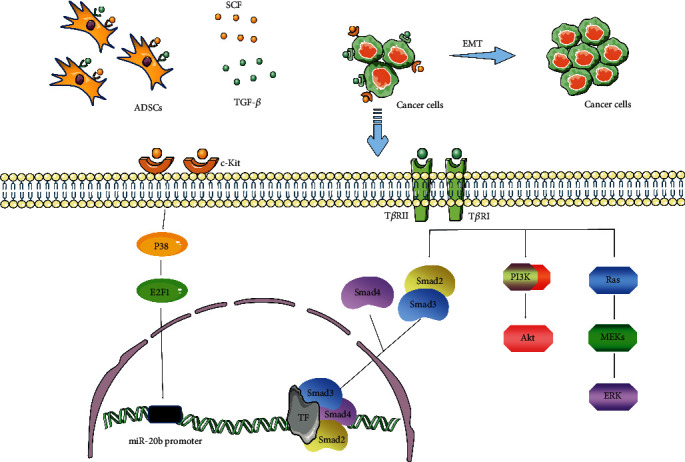
Adipose-derived stem cells (ADSCs) can maintain self-renewal and enhanced multidifferentiation potential through the release of a variety of paracrine factors and extracellular vesicles, allowing them to repair damaged organs and tissues. Consequently, considerable attention has increasingly been paid to their application in tissue engineering and organ regeneration. Here, we provide a comprehensive overview of the current status of ADSC preparation, including harvesting, isolation, and identification. The advances in preclinical and clinical evidence-based ADSC therapy for bone, cartilage, myocardium, liver, and nervous system regeneration as well as skin wound healing are also summarized. Notably, the perspectives, potential challenges, and future directions for ADSC-related researches are discussed. We hope that this review can provide comprehensive and standardized guidelines for the safe and effective application of ADSCs to achieve predictable and desired therapeutic effects.


Conflict of interest statement

The authors declare that there is no conflict of interest regarding the publication of this article.

Figures

Scheme 1
Scheme 1
Schematic representation of the applications for ADSC-based therapies in regenerative medicine. PDGF: platelet-derived growth factor; HGF: hepatocyte growth factor; IGF-1: insulin-like growth factor 1; BMP: bone morphogenetic protein; SCI: spinal cord injury; TBI: traumatic brain injury; ALF: acute liver failure; STEMI: ST-elevation acute myocardial infarction.
Figure 1
Figure 1
The typical process for the preparation of ADSCs from human adipose tissue. SVF: stromal vascular fraction.
Figure 2
Figure 2
Results of the expression of bone markers and mechanical properties of scaffold construct (a). Expression of osteopontin and collagen type I in sections of decalcified tibial samples (b). Locations of the indentation experiments on the empty hydroxyapatite disk (control) and the ADSC-hydroxyapatite disk (ADSCs) (c). The reduced modulus Er and hardness H values of the two groups at the three loads were investigated. Adapted from a previous study [58], with permission.
Figure 3
Figure 3
Flow chart of the experimental steps for long-term cartilage repair in rabbits (a). Immunohistochemical staining of interleukin- (IL-) 1β, IL-6, IL-10, and tumor necrosis factor (TNF). Black solid arrows denote the positive expression of IL-6 in the repair interface (b). Histological analysis of the cartilage defect after 3 and 6 months by hematoxylin and eosin (H&E) staining. Black solid arrows denote the repair interface. Red solid arrows denote the depth of the repaired cartilage (c). HC: host cartilage; RC: repaired cartilage. Adapted from a previous study [84], with permission.
Figure 4
Figure 4
Double immunostaining with anti-iNOS and anti-Iba-1 antibodies to identify M1 and M2 microglia in the cortex within 1 mm of the lesion in the sham, TBI, and TBI+secretome of ADSCs (TBI+ST) groups 7 days after traumatic brain injury (TBI) (a). Cytokine expression levels at 3 and 14 days after TBI were evaluated by qPCR (b, c). iNOS: inducible nitric oxide synthase; Iba-1: ionized calcium-binding adaptor molecule 1; Arg-1: arginase 1. Adapted from a previous study [119], with permission.
Figure 5
Figure 5
Representative images of semithin cross-sections of the regenerating sciatic nerve in the Dulbecco's modified Eagle's medium (DMEM) and ADSCs (a–c). Electron micrographs of a regenerating sciatic nerve in the transverse plane (b, c, e, f). Graph showing the total number of myelinated fibers in the sciatic nerve for all the groups (g). Quantitative morphological analyses of the axon area, fiber area, and myelin area in the regenerating sciatic nerve (h–j). Adapted from a previous study [132], with permission.
Figure 6
Figure 6
Scanning electron micrographs of Col-T gel-encapsulated ADSCs 3 days after encapsulation (a). Representative images of Masson trichrome staining of the transverse planes of heart sections (b). LVEF, LVESD, and LVEDD at 1 day, 2 weeks, and 4 weeks after myocardial infarction (c). LVEF: left ventricular ejection fraction; LVESD: left ventricular end-systolic diameter; LVEDD: left ventricular end-diastolic diameter. Adapted from a previous study [144], with permission.
Figure 7
Figure 7
A schematic representation of the experimental design (a). Scanning electron micrographs of ADSCs in a pEGFP-C1-transfected ADSCs/scaffolds and FOXA2-transfected ADSCs/scaffolds (b). Hematoxylin and eosin (H&E) staining of the necrotic area and retrieved scaffolds (c). TAA: thioacetamide. Adapted from a previous study [158], with permission.
Figure 8
Figure 8
Schematic showing how seawater (SW) and adipose-derived stem cells (ADSCs) regulate wound healing through the EGFR/MEK/ERK signaling pathway (a). The EGF protein expression levels were significantly higher in the control and SW+ADSC groups than in the SW and SW+DMEM groups (b). Hematoxylin and eosin (H&E) staining for wound repair, skin thickness, and a number of subcutaneous appendages (c). The red arrow denotes a hair follicle. Adapted from a previous study [165], with permission.
Figure 9
Figure 9
ADSCs promote the migration, invasion, and mesenchymal-epithelial transformation of cancer cells by secreting TGF-β and SCF. TF: transcription factor; TβR: TGF-β receptor.

References

    1. Rana A., Gruessner A., Agopian V. G., et al. Survival benefit of solid-organ transplant in the United States. JAMA Surgery. 2015;150(3):252–259. doi: 10.1001/jamasurg.2014.2038. - DOI - PubMed
    1. Jones B. Keeping kidneys. Bulletin of the World Health Organization. 2012;90(10):718–719. doi: 10.2471/BLT.12.021012. - DOI - PMC - PubMed
    1. Guess A. J., Daneault B., Wang R., et al. Safety profile of good manufacturing practice manufactured interferon γ-primed mesenchymal stem/stromal cells for clinical trials. Stem Cells Translational Medicine. 2017;6(10):1868–1879. doi: 10.1002/sctm.16-0485. - DOI - PMC - PubMed
    1. Carriel V., Geuna S., Alaminos M. Ex vivo and in vivo stem cells-based tissue engineering strategies for their use in regenerative medicine. Stem Cells International. 2018;2018:2. doi: 10.1155/2018/7143930.7143930 - DOI - PMC - PubMed
    1. Bhartiya D. Clinical translation of stem cells for regenerative medicine. Circulation Research. 2019;124(6):840–842. doi: 10.1161/CIRCRESAHA.118.313823. - DOI - PubMed

LinkOut - more resources