. 2020 Dec 7:2020:6697577.
doi: 10.1155/2020/6697577. eCollection 2020.

Exosomes Derived from Human Urine-Derived Stem Cells Inhibit Intervertebral Disc Degeneration by Ameliorating Endoplasmic Reticulum Stress

Affiliations

Exosomes Derived from Human Urine-Derived Stem Cells Inhibit Intervertebral Disc Degeneration by Ameliorating Endoplasmic Reticulum Stress

HongFei Xiang et al. Oxid Med Cell Longev. .

Abstract

Objective: This study is aimed at determining the effects of human urine-derived stem cell-derived exosomes (USCs-exos) on pressure-induced nucleus pulposus cell (NPC) apoptosis and intervertebral disc degeneration (IDD) and on the ERK and AKT signaling pathways.

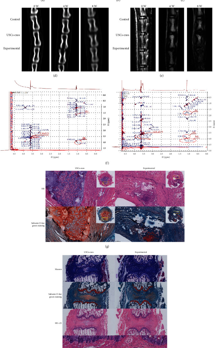
Methods: The NPCs were obtained from patients with herniated lumbar discs. Western blot analysis (WB) and quantitative real-time polymerase chain reaction (qRT-PCR) were used to determine endoplasmic reticulum (ER) stress levels of NPCs under stress. Human USCs were identified using an inverted microscope, three-line differentiation experiments, and flow cytometry. A transmission microscope, nanoparticle size analysis, and WB procedures were used to identify the extracted exosomes and observe NPC uptake. A control group, a 48 h group, and a USCs-exos group were established. The control group was untreated, and the 48 h group was pressure-trained for 48 h, while the USCs-exos group was pressure-trained for 48 h and treated with USCs-exos. WB, qRT-PCR, and terminal deoxynucleotidyl transferase dUTP nick end labeling (TUNEL) analysis were used to determine the ER stress levels in stress conditions and after exosomal treatment. The AKT and ERK pathways were partially detected. Magnetic Resonance Imaging (MRI) and computed tomography (CT) were used to evaluate cell degeneration while exosomal effects on the intervertebral disc (IVD) tissue were determined by hematoxylin and eosin (HE) staining, Safranin O-fast green staining, immunohistochemical staining (IHC), nuclear magnetic resonance (NMR), spectrometric detection, and total correlation spectroscopy (TOCSY). IVD metabolites were also identified and quantified.

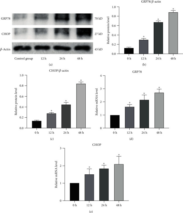
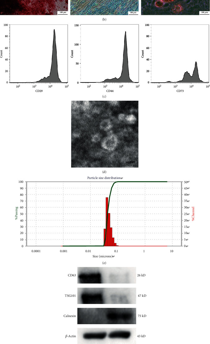
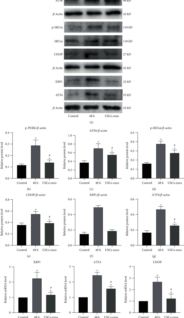
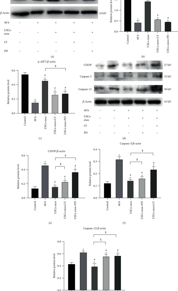
Results: After pressure culture, ER stress markers (GRP78 and C/EBP homologous protein (CHOP)) in the NPCs were significantly elevated with time (p < 0.05). Human USCs are short and spindle-shaped. They can successfully undergo osteogenic, adipogenic, and chondrogenic differentiation. In this study, these stem cells were found to be positive for CD29, CD44, and CD73. The exosomes were centrally located with a diameter of 50-100 nm. CD63 and Tsg101 were highly expressed while the expression of Calnexin was suppressed. The exosomes can be ingested by NPCs. USCs-exos significantly improved ER stress responses and inhibited excessive activation of the unfolded protein response (UPR) as well as cell apoptosis and disc degeneration through the AKT and ERK signaling pathways (p < 0.05).

Conclusion: Through the AKT and ERK signaling pathways, USCs-exos significantly inhibit ER stress-induced cell apoptosis and IDD under pressure conditions. It is, therefore, a viable therapeutic strategy.


Conflict of interest statement

The authors declare that they have no conflict of interest.

Figures

Figure 1
Figure 1
Endoplasmic reticulum stress level after stress culture of human NPCs. (a–c) Analysis of the protein levels of GRP78 and CHOP by Western blot (a), and based on this, the gray values of relative protein expression are compared (b, c). β-Actin was used as an internal control. Compared to the control group, p < 0.05. (d, e) The mRNA levels of GRP78 and CHOP in NPCs after pressure culture. Compared to the NC group, p < 0.05.
Figure 2
Figure 2
Identification of human USC exosomes (USCs-exos). (a) Human USCs exhibit a short fusiform or spindle-shaped appearance, with sporadic cells appearing on day three (left picture) and large numbers of cells appearing at day seven (right picture). (b) Osteogenic, adipogenic, and chondrogenic differentiation capabilities of USCs were determined by Alizarin Red, Oil Red O, and Alcian Blue staining. (c) USC surface markers (CD29, CD44, and CD73) were detected by flow cytometry. (d) A typical image of USCs-exos morphology as obtained by a transmission electron microscope (TEM). (e) Particle size distribution of USCs-exos as determined by the nanoparticle size analysis. (f) The protein marker of USCs-exos as detected in exosomes and USCs by Western blot analysis. (g) Exosomes taken by NP cells incubated with PKH26-labeled USCs-exos for 12 hours, and NP cell nuclei were stained with DAPI.
Figure 3
Figure 3
Under stress conditions, USCs-exos suppressed the expression of GRP78 and GRP94. Except the control group, NP cells were cultured for 48 h under pressure. USCs-exos-10, 50, and 100 indicated that 10, 50, or 100 μg/ml exosomes were added to each response group. (a) Protein levels of GRP78 and GRP94 were measured by Western blot analysis, and their relative quantities were calculated (b, c) using β-actin as an internal reference. (d) Western blot analysis was used to detect the expression levels of caspase-3 and caspase-12, and their relative quantities (e, f) were calculated using β-actin as an internal reference. (g) Fluorescence images of TUNEL analysis in different groups. The nucleus was stained with DAPI. (h) The proportion of apoptotic cells according to TUNEL staining. Data are expressed as the mean ± SD. Compared to the control group, p < 0.05; #compared to the 48 h group, p < 0.05.
Figure 4
Figure 4
USCs-exos enhance the activation of UPR and related proteins under stress conditions. The USCs-exos group was treated with USCs-exos (100 μg/ml). (a–g) Protein expression levels of p-PERK, PERK, ATF6, p-IRE1α, IRE1α, XBP1, ATF4, and CHOP as determined by Western blot analysis and calculation of their relative quantities (b–g) using β-actin as an internal reference. (h–j) The transcription levels of XBP1 (h), ATF4 (i), and CHOP (j) as determined by qRT-PCR. The data are expressed as the mean ± SD. Compared to the control group, p < 0.05; #compared to the 48 h group, p < 0.05.
Figure 5
Figure 5
USCs-exos regulate ER stress under stress-induced conditions by activating the AKT and ERK signaling pathways in NPCs. In the USCs-exos group, USCs-exos (100 μg/ml) was added for intervention under pressure. (a–c) Protein levels of AKT, p-AKT, ERK, and p-ERK were evaluated by Western blotting, and their relative quantities were calculated (b, c) using β-actin as the internal reference. LY294002 (LY) is an inhibitor of PI3K/AKT. PD98059 (PD) is an inhibitor of ERK1/2 phosphorylation. (d–g) The protein levels of CHOP, caspase-12, and caspase-3 were measured by Western blotting and statistically analyzed (e–g) using β-actin as an internal control. The data are expressed as the mean ± SD. Compared to the control group, p < 0.05; #compared to the 48 h group, p < 0.05; $compared to USCs-exos group, p < 0.05.
Figure 6
Figure 6
USCs-exos inhibits ER stress-associated cell apoptosis and delays IDD in vivo. (a–c) The expression levels of GRP78 and CHOP in rat degeneration models as determined by Western blotting, and their relative quantities (b, c). (d) CT scans of the three adjacent IVDs of the rat's tail vertebra at 0, 4, and 8 weeks. The height of the intervertebral space in the experimental group was significantly lower than that of the USCs-exos group and the control group. (e) T2W1-weighted images of the MRI scans performed on the rat's tail at 0, 4, and 8 weeks. The degeneration of the experimental group was significantly stronger than that of the USCs-exos group and control group. (f) In the NMR detection, compared to the simple puncture, the CHOP amino acid residue leucine (Leu) in the IVDs punctured and injected with USCs-exos was significantly suppressed; caspase-3 amino acid residue aspartic acid. The content of the injected exosomes in the IVD is significantly lower than that of the pure puncture segment; lactic acid (Lac) levels were also significantly low. (g) The HE and Safranin O-fast green staining of the IVD revealed that the IVD with simple puncture was more disordered and looser than the annulus fibrosus injected with USCs-exos and contained a large number of inflammatory cells and scars. The degenerated nucleus pulposus tissue protrudes into the annulus fibrosus. (h) Masson staining, Safranin O-fast green staining, and HE staining all showed that the degree of degeneration of the IVD tissue of injected USCs-exos was less, and IHC showed that the expression of caspase-3 was lower. Compared to the control group, p < 0.05; #compared to the USCs-exos group, p < 0.05.

Similar articles

Cited by

References

    1. Macfarlane G. J., Thomas E., Croft P. R., Papageorgiou A. C., Jayson M. I. V., Silman A. J. Predictors of early improvement in low back pain amongst consulters to general practice: the influence of pre-morbid and episode-related factors. Pain. 1999;80(1):113–119. doi: 10.1016/S0304-3959(98)00209-7. - DOI - PubMed
    1. Fu J., Yu W., Jiang D. Acidic pH promotes nucleus pulposus cell senescence through activating the p38 MAPK pathway. Bioscience Reports. 2018;38(6) doi: 10.1042/bsr20181451. - DOI - PMC - PubMed
    1. Frost B. A., Camarero-Espinosa S., Foster E. J. Materials for the spine: anatomy, problems, and solutions. Materials. 2019;12(2):p. 253. doi: 10.3390/ma12020253. - DOI - PMC - PubMed
    1. ZHANG F., ZHAO X., SHEN H., ZHANG C. Molecular mechanisms of cell death in intervertebral disc degeneration (review) International Journal of Molecular Medicine. 2016;37(6):1439–1448. doi: 10.3892/ijmm.2016.2573. - DOI - PMC - PubMed
    1. Ding F., Shao Z. W., Xiong L. M. Cell death in intervertebral disc degeneration. Apoptosis. 2013;18(7):777–785. doi: 10.1007/s10495-013-0839-1. - DOI - PubMed