. 2020 Dec 31:2020:8706898.
doi: 10.1155/2020/8706898. eCollection 2020.

HSP60 Regulates Monosodium Urate Crystal-Induced Inflammation by Activating the TLR4-NF- κ B-MyD88 Signaling Pathway and Disrupting Mitochondrial Function

Affiliations

HSP60 Regulates Monosodium Urate Crystal-Induced Inflammation by Activating the TLR4-NF- κ B-MyD88 Signaling Pathway and Disrupting Mitochondrial Function

Qiushi Huang et al. Oxid Med Cell Longev. .

Abstract

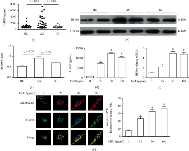
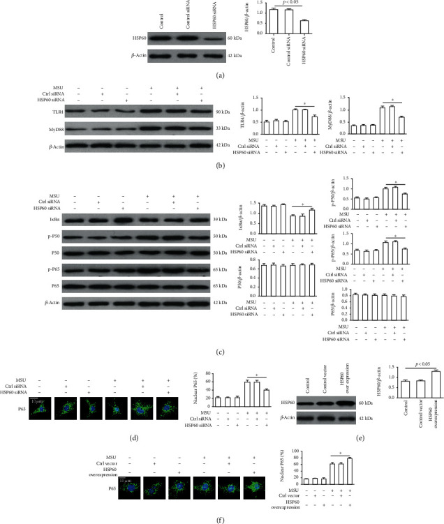
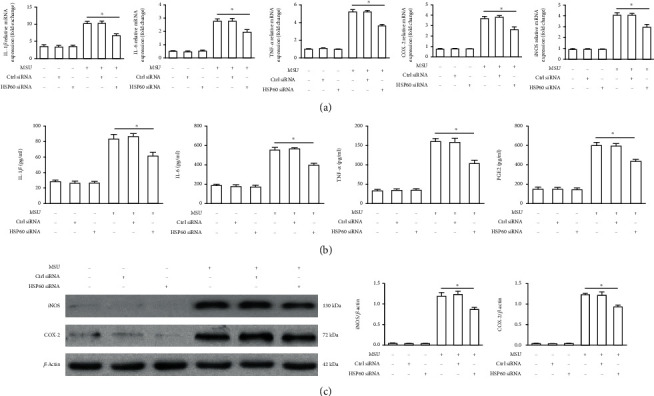
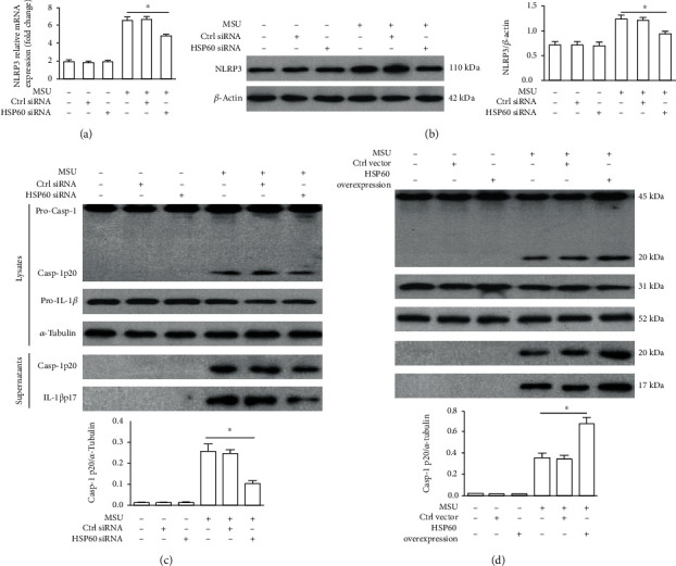
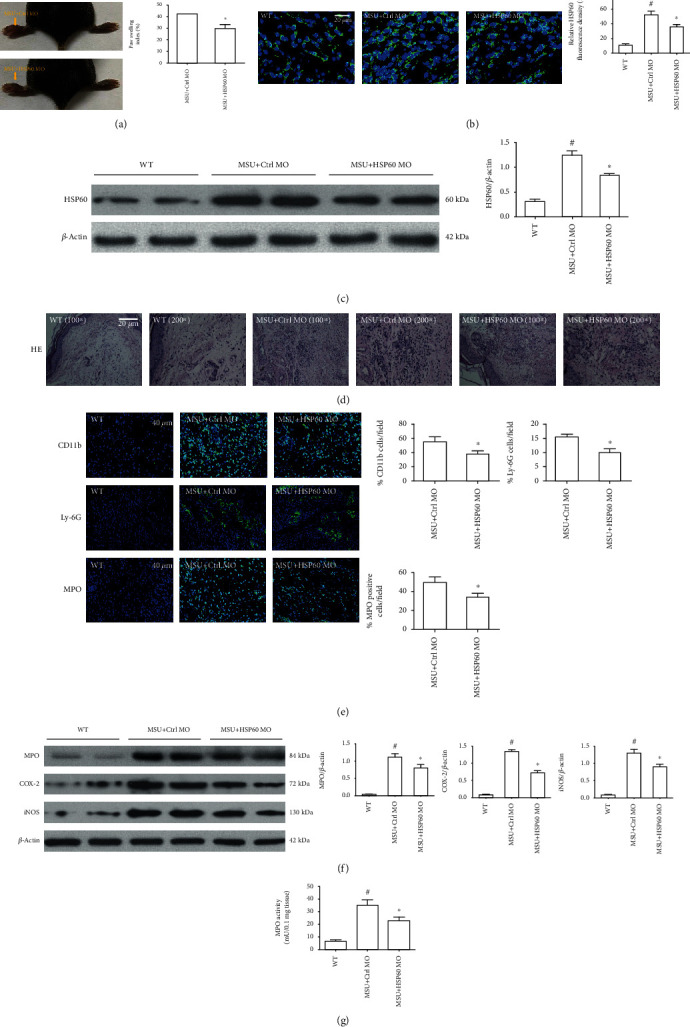
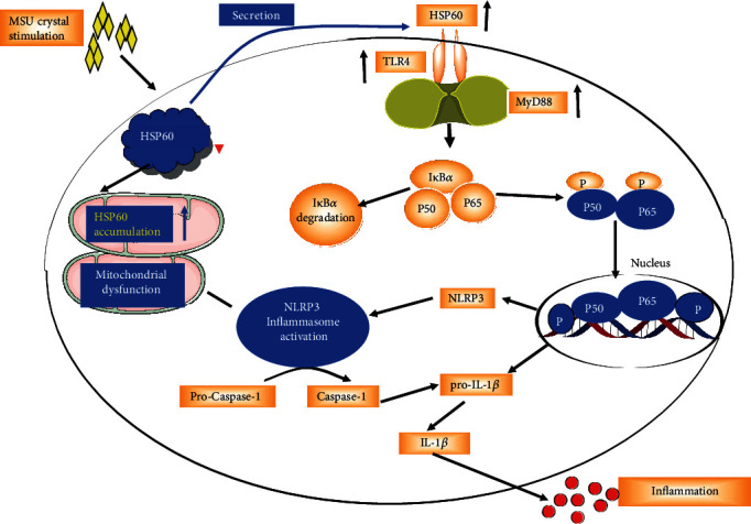
Acute gout is an inflammatory response induced by monosodium urate (MSU) crystals. HSP60 is a highly conserved stress protein that acts as a cellular "danger" signal for immune reactions. In this study, we aimed to investigate the role and molecular mechanism of HSP60 in gout. HSP60 expression was detected in peripheral blood mononuclear cells (PBMCs) and plasma of gout patients. The effect and molecular mechanism of HSP60 in gout were studied in MSU crystals treatment macrophages and C57BL/6 mice. JC-1 probe and MitoSOX Red were used to measure the mitochondrial membrane potential (MMP) and mitochondrial reactive oxygen species (mtROS). HSP60 expression was significantly upregulated in the PBMCs and sera of patients with acute gout (AG) compared to those with intercritical gout (IG) or healthy controls (HCs). MSU crystals induced the expression and secretion of HSP60 in the macrophages. HSP60 knockdown or overexpression affects TLR4 and MyD88 expression, IκBα degradation, and the nuclear localization of NF-κB in MSU crystal-stimulated inflammation. Further, HSP60 facilitates MMP collapse and mtROS production and activates the NLRP3 inflammasome in MSU crystal-stimulated macrophages. In MSU crystal-induced arthritis mouse models pretreated with HSP60 vivo-morpholino, paw swelling, myeloperoxidase (MPO) activity, and inflammatory cell infiltration significantly decreased. Our study reveals that MSU crystal stimulates the expression of HSP60, which accelerates the TLR4-MyD88-NF-κB signaling pathway and exacerbates mitochondrial dysfunction.


Conflict of interest statement

The authors declare no potential conflicts of interest.

Figures

Figure 1
Figure 1
HSP60 expression was upregulated in gout and MSU crystal-stimulated THP-1-derived macrophages. (a) HSP60 protein level in the plasma from patients with AGA (n = 31), IGA (n = 33), and HC (n = 43) was detected by ELISA. (b) HSP60 protein level in PBMCs of patients with AGA, IGA, and HCs was measured by western blot (8 cases in each group). (c) Densitometry analysis of HSP60 protein level in PBMCs. (d–f) THP-1-derived macrophages were primed with LPS (100 ng/ml) for 1 h and then treated with different concentrations of MSU suspension for 12 h. (d) HSP60 secretion was analyzed by ELISA in THP-1-derived macrophages. (e) The relative mRNA expression of HSP60 in MSU crystal-stimulated THP-1-derived macrophages. (f) Representative images of double-labeling HSP60 and mitochondrial marker (MitoTracker) in MSU crystal-stimulated THP-1-derived macrophages. Scale bar: 20 μm. Blue shows nuclei staining with DAPI, quantification of immunofluorescence staining of HSP60 using Image J software. In comparison with absence of MSU crystal treatment and # in comparison with 25 μg/ml MSU crystal treatment.
Figure 2
Figure 2
HSP60 affects the TLR4/MyD88/NF-κB signaling pathway in MSU crystal-stimulated RAW264.7 cells. After RAW264.7 cells were transfected with HSP60 siRNA or control siRNA for 48 h, primed with LPS (100 ng/ml) for 1 h, and then treated with MSU crystals (100 μg/ml) for 4 h. (a) HSP60 protein level in RAW264.7 cells transfected with ctrl siRNA or HSP60 siRNA. (b, c) Protein levels of TLR4, MyD88, IκBα, P50, P65, phosphorylated P50 (p-P50), and phosphorylated P65 (p-P65). (d) HSP60 knockdown inhibited MSU crystal-induced P65 nuclear localization in RAW264.7 cells and analyzed by immunostaining. Blue shows nuclei staining with DAPI. Scale bar: 10 μm. Each experiment had six fields of view. The intensity of the fluorescence signal was analyzed by Image J software. (e) HSP60 protein level in RAW264.7 cell transfection with control vector or HSP60 vector for 36 h. (f) RAW264.7 cells were transfected with a control vector or HSP60 vector for 36 h, primed with LPS (100 ng/ml) for 1 h, primed with LPS (100 ng/m), and then treated with MSU crystals (100 μg/ml) for 4 h. HSP60 overexpression promoted MSU crystal-induced P65 nuclear localization in RAW264.7 cells and analyzed by immunostaining. Blue shows nuclei staining with DAPI. Scale bar: 10 μm. Each experiment had six fields of view. The intensity of the fluorescence signal was analyzed by Image J software. Values are the mean ± SEM of 3 independent experiments. p < 0.05.
Figure 3
Figure 3
HSP60 knockdown reduced the expression of MSU crystal-induced inflammatory associated gene in THP-1-derived macrophages. THP-1-derived macrophages were transfected with ctrl siRNA or HSP60 siRNA for 48 h, primed with LPS (100 ng/ml) for 1 h, and then stimulated with MSU crystals (50 μg/ml) for 12 h. (a) Quantification of RT-PCR analysis of IL-1β, IL-6, TNFα, COX-2, and iNOS mRNA expression. (b) The release of IL-1β, IL-6, TNFα, and PGE2 in the culture supernatant was measured by ELISA. (c) Western blot analysis of COX-2 and iNOS protein levels. Data are represented as the mean ± SEM for three experiments. p < 0.05.
Figure 4
Figure 4
HSP60 affects the activation of the NLRP3 inflammasome in MSU crystal-treated THP-1-derived macrophages. (a–c) THP-1-derived macrophages were transfected with control siRNA or HSP60 siRNA for 48 h, primed with LPS (100 ng/ml) for 1 h, and then treated with MSU suspension (50 μg/ml) for 12 h. (a) Effect of HSP60 knockdown on NLRP3 mRNA levels. (b) Influence effect of HSP60 knockdown on NLRP3 protein levels. (c) Effect of HSP60 knockdown on the expression of the p20 subunit of Caspase-1 and IL-1β in the cell lysates and culture supernatants. (d) THP-1-derived macrophages were transfected with a control vector or human HSP60 vector for 36 h, primed with LPS (100 ng/ml) for 1 h, and then treated with MSU suspension (50 μg/ml) for 12 h. Effect of HSP60 overexpression on the expression of the p20 subunit of Caspase-1 and IL-1β in the cell lysates and culture supernatants. The p20 subunit of Caspase-1 protein levels in the cell lysates was normalized to ɑ-Tubulin and then averaged. Values are the mean ± SEM of 3 independent experiments. p < 0.05.
Figure 5
Figure 5
HSP60 knockdown alleviates mitochondrial dysfunction in MSU crystal-stimulated THP-1-derived macrophages. (a–g) THP-1-derived macrophages were transfected with control siRNA or HSP60 siRNA for 48 h, primed with LPS (100 ng/ml) for 1 h, and then treated with MSU suspension (50 μg/ml) for 12 h. (a) The effect of HSP60 knockdown on the mitochondrial ROS, representative images of MitoTracker green and MitoROS staining. Blue shows nuclei staining with Hoechst33342. Scale bar: 20 μm. (b) SOD activity. (c) CAT activity. (d) GSH-Px activity. (e) Mitochondrial DNA release was detected by quantitative real-time PCR analysis. (f) Relative mitochondrial ATP content. (g) The effect of HSP60 knockdown on the mitochondrial membrane potential (MMP), cells were stained using JC-1 probe. Blue shows nuclei staining with Hoechst33342. Scale bar: 20 μm. For mtROS and MMP analysis, >50 individual cells were imaged per group from 3 culture dishes. Data are representative of mean ± SEM for three experiments. Each experiment had 10 fields of view. p < 0.05.
Figure 6
Figure 6
HSP60 downregulation relieved the severity of MSU crystal-induced inflammatory mouse model. (a) Mice injected with HSP60-MO presented decreased the paw swelling index (n = 6 per group, mean ± SEM). (b) Immunostaining was used to detect HSP60 expression in the foot pad tissue section (n = 4 per group, mean ± SEM). Blue shows nuclei staining with DAPI. Scale bar: 20 μm. (c) HSP60 protein levels were analyzed by western blot in the foot pad tissue (n = 4 per group, mean ± SEM). (d) HE staining was used to observe the infiltration of inflammatory cells in the foot pad tissue sections (n = 4 per group, mean ± SEM). Images were obtained using a light microscope (Olympus, Tokyo, Japan) with 10 times or 20 times objectives and analyzed with image processing software (Olympus). (e) Immunostaining using anti-CD11b, anti-Ly-6G, and anti-MPO antibody in the foot pad tissue sections (n = 4 per group, mean ± SEM). Blue shows nuclei staining with DAPI. Scale bar: 40 μm. (f) Protein levels of MPO, iNOS, and COX-2 in the foot pad tissue were measured by western blot (n = 4 per group, mean ± SEM). (g) The MPO activity of foot pad tissue (n = 6 per group, mean ± SEM). #Significantly different from the absence of MSU crystals injection mice and significantly different from MSU crystals and ctrl MO injection mice.
Figure 7
Figure 7
Schema of the signaling pathway involved in HSP60-mediated NLRP3 inflammasome activation and subsequent IL-1β production. After MSU crystal stimulation, the secretion of HSP60 to the extracellular level is greatly increased, which activates the signaling pathway and promotes the transcription of IL-1β and NLRP3. MSU crystal treatment also leads to a large amount of HSP60 aggregation in mitochondria, causing mitochondrial dysfunction, thereby activating NLRP3 inflammasome and promoting IL-1β precursor processing and ultimately leading to IL-1β release and inflammatory response.

Similar articles

Cited by

References

    1. Chen‐Xu M., Yokose C., Rai S. K., Pillinger M. H., Choi H. K. Contemporary prevalence of gout and hyperuricemia in the United States and decadal trends: the National Health and Nutrition Examination Survey, 2007-2016. Arthritis & Rheumatology. 2019;71(6):991–999. doi: 10.1002/art.40807. - DOI - PMC - PubMed
    1. Shi Y., Evans J. E., Rock K. L. Molecular identification of a danger signal that alerts the immune system to dying cells. Nature. 2003;425(6957):516–521. doi: 10.1038/nature01991. - DOI - PubMed
    1. Zhang S., Zhang Y., Liu P., Zhang W., Ma J.-l., Wang J. Efficacy and safety of etoricoxib compared with NSAIDs in acute gout: a systematic review and a meta-analysis. Clinical Rheumatology. 2016;35(1):151–158. doi: 10.1007/s10067-015-2991-1. - DOI - PubMed
    1. Richette P., Doherty M., Pascual E., et al. 2016 updated EULAR evidence-based recommendations for the management of gout. Annals of the Rheumatic Diseases. 2017;76(1):29–42. doi: 10.1136/annrheumdis-2016-209707. - DOI - PubMed
    1. Rossato M. F., Hoffmeister C., Trevisan G., et al. Monosodium urate crystal interleukin-1β release is dependent on Toll-like receptor 4 and transient receptor potential V1 activation. Rheumotology. 2020;59(1) doi: 10.1093/rheumatology/kez384. - DOI - PubMed

MeSH terms