. 2020 Dec 7:2020:8862953.
doi: 10.1155/2020/8862953. eCollection 2020.

What Is the Impact of Depletion of Immunoregulatory Genes on Wound Healing? A Systematic Review of Preclinical Evidence

Affiliations

What Is the Impact of Depletion of Immunoregulatory Genes on Wound Healing? A Systematic Review of Preclinical Evidence

Bárbara Cristina Félix Nogueira et al. Oxid Med Cell Longev. .

Abstract

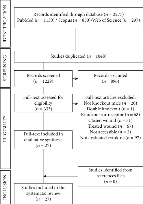
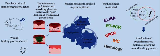
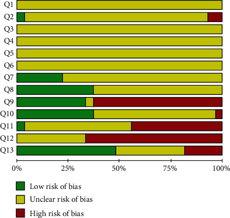
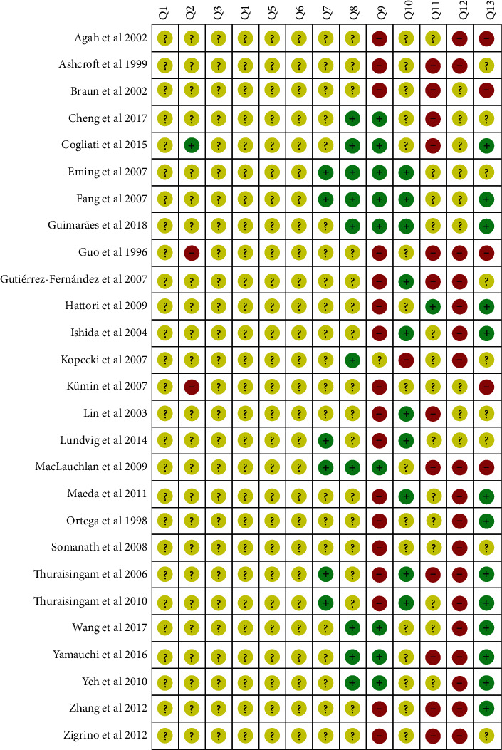
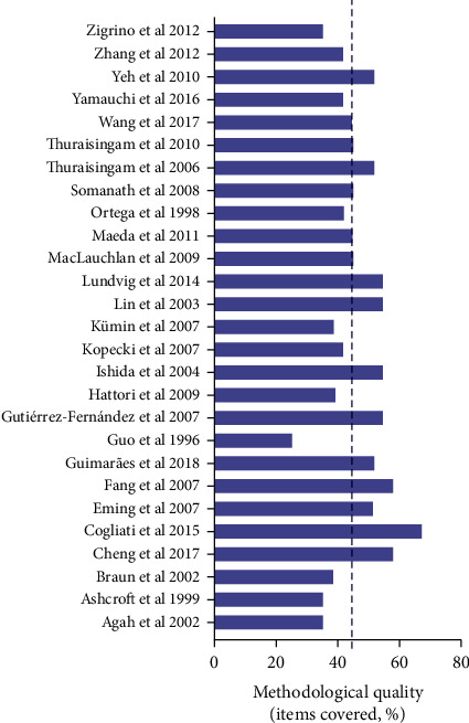
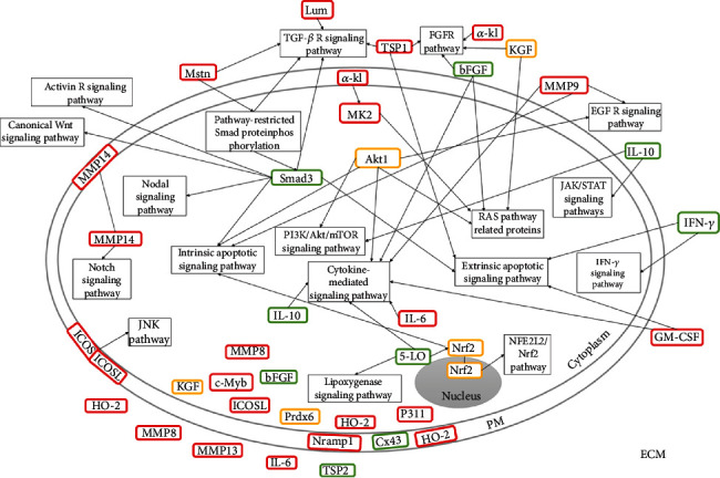
Cytokines and growth factors are known to play an important role in the skin wound closure process; however, in knockout organisms, the levels of these molecules can undergo changes that result in the delay or acceleration of this process. Therefore, we systematically reviewed evidence from preclinical studies about the main immunoregulatory molecules involved in skin repair through the analysis of the main mechanisms involved in the depletion of immunoregulatory genes, and we carried out a critical analysis of the methodological quality of these studies. We searched biomedical databases, and only original studies were analyzed according to the PRISMA guidelines. The included studies were limited to those which used knockout animals and excision or incision wound models without intervention. A total of 27 studies were selected; data for animal models, gene depletion, wound characteristics, and immunoregulatory molecules were evaluated and compared whenever possible. Methodological quality assessments were examined using the ARRIVE and SYRCLE's bias of risk tool. In our review, the extracellular molecules act more negatively in the wound healing process when silenced and the metabolic pathway most affected involved in these processes was TGF-β/Smad, and emphasis was given to the importance of the participation of macrophages in TGF-β signaling. Besides that, proinflammatory molecules were more evaluated than anti-inflammatory ones, and the main molecules evaluated were, respectively, TGF-β1, followed by VEGF, IL-6, TNF-α, and IL-1β. Overall, most gene depletions delayed wound healing, negatively influenced the concentrations of proinflammatory cytokines, and consequently promoted a decrease of inflammatory cell infiltration, angiogenesis, and collagen deposition, compromising the formation of granulation tissue. The studies presented heterogeneous data and exhibited methodological limitations; therefore, mechanistic and highly controlled studies are required to improve the quality of the evidence.


Conflict of interest statement

The authors declare that there are no conflicts of interest.

Figures

Figure 1
Figure 1
PRISMA diagram. Different phases of the selection of studies for conducting qualitative and quantitative analyses. Flow diagram of the systematic review literature search results. Based on “Preferred Reporting Items for Systematic Reviews and Meta-Analyses: The PRISMA Statement.” http://www.prisma-statement.org. From: Moher D, Liberati A, Tetzlaff J, Altman DG, The PRISMA Group (2009).
Figure 2
Figure 2
A schematic diagram showing the influence of different immunoregulatory genes of knockout mice on the wound healing process, the main phases affected by genetic silencing, and consequently the main mechanisms involved in this process, and the most common techniques used for this type of analysis. KO: Knockout; ELISA: Enzyme-Linked Immunosorbent Assay; RT-PCR: Reverse Transcription Polymerase Chain Reaction; qPCR: Real-Time quantitative Polymerase Chain Reaction; IHC: Immunohistochemistry; TGF-β1: Transforming Growth Factor beta 1; VEGF: Vascular Endothelial Growth Factor; TNF-α: Tumor Necrosis Factor-alpha; IL-6: Interleukin-6; IL-1β: Interleukin-1 beta; ep: epidermis; dm: dermis; inf: inflammatory molecules.
Figure 3
Figure 3
Bias risk results and methodological quality indicators for all studies included in this systematic review that evaluated the effect of gene depletion on excisional and incisional wounds. Q1: Was the allocation sequence adequately generated and applied? Q2: Were the groups similar at baseline or were they adjusted for confounders in the analysis? Q3: Was the allocation to the different groups adequately concealed? Q4: Were the animals randomly housed during the experiment? Q5: Were the caregivers and/or investigators blinded from knowledge regarding which intervention each animal received during the experiment? Q6: Were animals selected at random for outcome assessment? Q7: Was the outcome assessor-blinded? Q8: Were incomplete outcome data adequately addressed? Q9: Are reports of the study free of selective outcome reporting? Q10: Was the study free of other problems that could result in a high bias risk? Q11: Was the number of animals per group and number of animals per cage presented? Q12: What conditions were the animals kept in? and Q13: Wound closure data were presented with follow-up days, photos, and graphs?
Figure 4
Figure 4
Risk of bias summary: review authors' judgments about the risk of bias items for each included study. Green: low risk of bias; Yellow: unclear risk of bias; and Red: high risk of bias.
Figure 5
Figure 5
Analysis of methodological bias (reporting quality) for each study included in the review. Based on Animal Research: Reporting of In Vivo Experiments (ARRIVE) guidelines (http://www.nc3rs.org.uk/arrive-guidelines). The dotted line indicated the mean quality score (%). Detailed bias analysis stratified by domains and items evaluated is presented in Table S3.
Figure 6
Figure 6
Location of the molecules of depleted genes addressed in this review and their participation in metabolic pathways involved in the wound repair. The effect of depleted genes on wound closure was shown by the colors: red (delayed), green (accelerated), and yellow (unchanged). PM: plasma membrane; ECM: extracellular matrix; 5-LO: 5-Lipoxygenase; α-kl: alpha-Klotho; bFGF: basic Fibroblastic Growth Factor; Cx43: Connexin 43; GM-CSF: Granulocyte-Macrophage Colony-Stimulating Factor; HO-2: Heme Oxygenase 2; ICOS: Inducible Costimulator; ICOSL: Inducible Costimulator Ligand; IFN-γ: Interferon-gamma; (IL-10, IL-6): Interleukins; KGF: Keratinocyte Growth Factor; Lum: Lumican; (MMP8, MMP9, MMP13, MMP14): Matrix Metalloproteinase; MK2: Mitogen-Activated Protein Kinase-2; Mstn: Myostatin; Nramp: Natural resistance-associated macrophage proteins; P311: Neuronal protein 3.1; Prdx6: Peroxiredoxin 6; Akt1: Serine/threonine kinase; (TPS1, TPS2): Thrombospondin; Nrf2: Transcription factor NF-E2-related factor 2; c-Myb: Transcription factor proto-oncogene c-Myb. Signaling pathways: Activin R: Activin Receptor; FGF R: Fibroblast Growth Factor Receptor; EGF R: Epidermal Growth Factor Receptor; TGF-β R: Transforming Growth Factor-β Receptor; NFE2L2/Nrf2: Nuclear Factor, Erythroid 2 Like 2; JNK: c-Jun N-terminal kinase; IFN-γ: Interferon-gamma; JAK/STAT: Janus Kinase Signal Transducer and Activator of Transcription.

Similar articles

Cited by

References

    1. Sen C. K., Gordillo G. M., Roy S., et al. Human skin wounds: a major and snowballing threat to public health and the economy. Wound Repair and Regeneration. 2009;17(6):763–771. doi: 10.1111/j.1524-475X.2009.00543.x. - DOI - PMC - PubMed
    1. Hingorani A., LaMuraglia G. M., Henke P., et al. The management of diabetic foot: a clinical practice guideline by the Society for Vascular Surgery in collaboration with the American Podiatric Medical Association and the Society for Vascular Medicine. Journal of Vascular Surgery. 2016;63(2):3S–21S. doi: 10.1016/j.jvs.2015.10.003. - DOI - PubMed
    1. Peter-Riesch B. The diabetic foot: the never-ending challenge. Novelties in Diabetes; - DOI - PubMed
    1. Schaper N. C., Van Netten J. J., Apelqvist J., Lipsky B. A., Bakker K. Prevention and management of foot problems in diabetes: a Summary Guidance for Daily Practice 2015, based on the IWGDF Guidance Documents. Diabetes/Metabolism Research and Reviews. 2016;32:7–15. doi: 10.1002/dmrr.2695. - DOI - PubMed
    1. Newbern S. Identifying pain and effects on quality of life from chronic wounds secondary to lower-extremity vascular disease. Advances in Skin & Wound Care. 2018;31(3):102–108. doi: 10.1097/01.ASW.0000530069.82749.e5. - DOI - PubMed

Publication types

LinkOut - more resources