. 2020 Dec 31:2020:9782062.
doi: 10.1155/2020/9782062. eCollection 2020.

Neuroprotection Effect of Astragaloside IV from 2-DG-Induced Endoplasmic Reticulum Stress

Affiliations

Neuroprotection Effect of Astragaloside IV from 2-DG-Induced Endoplasmic Reticulum Stress

Yu Fu et al. Oxid Med Cell Longev. .

Abstract

Objective: Astragaloside IV shows neuroprotective activity, but its mechanism remains unclear. To investigate whether astragaloside IV protects from endoplasmic reticulum stress (ERS), we focus on the regulation of glycogen synthase kinase-3β (GSK-3β) and mitochondrial permeability transition pore (mPTP) by astragaloside IV in neuronal cell PC12.

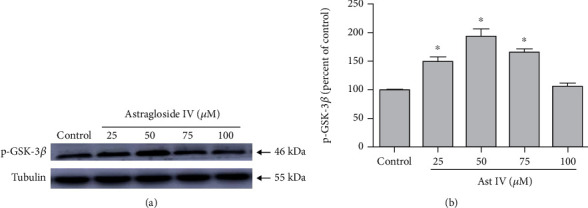
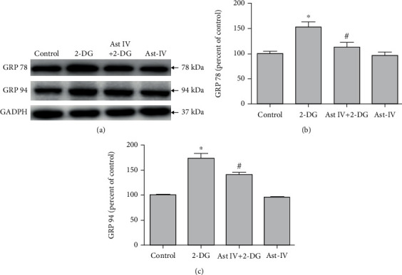
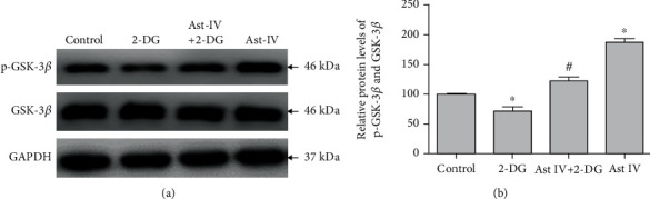
Methods and results: PC12 cells treated with different concentrations of ERS inductor 2-deoxyglucose (2-DG) (25-500 μM) showed a significant increase of glucose-regulated protein 78 (GRP 78) and GRP 94 expressions and a decrease of tetramethylrhodamine ethyl ester (TMRE) fluorescence intensity and mitochondrial membrane potential (∆Ψm), with the peak effect seen at 50 μM, indicating that 2-DG induces ERS and the mPTP opening. Similarly, 50 μM of astragaloside IV increased the GSK-3β phosphorylation at Ser9 most significantly. Next, we examined the neuroprotection of astragaloside IV by dividing the PC12 cells into control group, 2-DG treatment group, astragaloside IV plus 2-DG treatment group, and astragaloside IV only group. PC12 cells treated with 50 μM 2-DG for different time courses (0-36 hr) showed a significant increase of Cleaved-Caspase-3 with the peak at 6 hr. 2-DG significantly induced cell apoptosis and increased the green fluorescence intensity of Annexin V-FITC, and these effects were reversed by astragaloside IV. Such a result indicates that astragaloside IV protected neural cell survival from ERS. 2-DG treatment significantly increased the expressions of inositol-requiring ER-to-nucleus signal kinase 1 (IRE1), phosphor-protein kinase R-like ER kinase (p-PERK), but not affect the transcription factor 6 (ATF6) expression. 2-DG treatment significantly decreased the phosphorylation of GSK-3β and significantly reduced the TMRE fluorescence intensity and ∆Ψm, following mPTP open. Astragaloside IV significantly inhibited the above effects caused by 2-DG, except the upregulation of ATF6 protein. Taken together, astragaloside IV significantly inhibited the ERS caused by 2-DG.

Conclusion: Our data suggested that astragaloside IV protects PC12 cells from ERS by inactivation of GSK-3β and preventing the mPTP opening. The GRP 78, GRP 94, IRE1, and PERK signaling pathways but not ATF6 are responsible for GSK-3β inactivation and neuroprotection by astragaloside IV.


Conflict of interest statement

The authors declare no conflict of interest associated with this manuscript.

Figures

Figure 1
Figure 1
Effects of 2-DG on ERS and the mPTP opening. (a) Western blot images of GRP 78 and GRP 94 expression in PC12 cells treated with vehicle control and 25, 50, 100, 150, 200, and 500 μM 2-DG. (b) Confocal fluorescence images of cells incubating TMRE for 20 minutes in PC12 cells treated with 0, 25, 50, 100, 150, 200, and 500 μM 2-DG. (c, d) Quantification of (a). Each protein expression level was normalized with GAPDH. (e) Quantification of (b). Data are mean ± SD of 6 independent experiments. P < 0.05 compared to vehicle control. Scale bar, 50 μm.
Figure 2
Figure 2
Effect of astragaloside IV on GSK-3β phosphorylation. (a) Western blot image of p-GSK-3β expression in PC12 cells treated with vehicle control and 25, 50, 75, and 100 μM astragaloside IV. (b) Quantification of (a). Each protein expression level was normalized with Tubulin. Data are mean ± SD of 6 independent experiments.P < 0.05 compared to vehicle control.
Figure 3
Figure 3
Effect of astragaloside IV on PC12 cell survival from ERS. (a) Representative Western blot images of Cleaved-Caspase-3 in PC12 cells treated with 2-DG in different times. (b) Representative Western blot images of Cleaved-Caspase-3 in PC12 cells treated with 2-DG and astragaloside IV. (c, d) Confocal fluorescence images and flow cytometry of Annexin V-FITC Apoptosis Detection kit in PC12 cells treated with 2-DG and astragaloside IV. Cells were preloaded with Annexin V and PI for 20 minutes before other treatments. PC12 cell culture was placed on the temperature-controlled stage and treated with astragaloside IV for 20 minutes and 2-DG for 6 h. Data are mean ± SD of 6 independent experiments. P < 0.05 compared to control; #P < 0.05 compared to 2-DG. Scale bar, 50 μm.
Figure 4
Figure 4
Effect of astragaloside IV on PC12 cells from ERS. (a) Western blot images of ER chaperone proteins GRP 78 and GRP 94 in PC12 cells treated with 2-DG and astragaloside IV. (b, c) Quantification of (a). Each protein expression level was normalized with GAPDH. Data are mean ± SD of 6 independent experiments. P < 0.05 compared to vehicle control; #P < 0.05 compared to 2-DG.
Figure 5
Figure 5
Effect of astragaloside IV on PC12 cells from ERS-induced mPTP opening. (a) Confocal images of TMRE probe in PC12 cells treated with 2-DG and astragaloside IV. Cells were preloaded with TMRE probe for 20 minutes before other treatments. PC12 cell culture was placed on the temperature-controlled stage and treated with astragaloside IV for 20 minutes and 2-DG for 30 minutes. (b) Quantification of TMRE Red fluorescence expressed as % of the control. The fluorescence intensity value was calculated as 30-minute normalized to 0-minute. Data are mean ± SD of 6 independent experiments. P < 0.05 compared to control; #P < 0.05 compared to 2-DG. Scale bar, 50 μm.
Figure 6
Figure 6
Effect of astragaloside IV on ERS signaling pathway. (a) Representative Western blot images of IRE1, p-PERK, and ATF6 in PC12 cells treated with 2-DG and astragaloside IV. (b–d) Quantification of (a). Each protein expression level was normalized with GAPDH. Data are mean ± SD for 6 independent experiments. P < 0.05 compared to vehicle control; #P < 0.05 compared to 2-DG.
Figure 7
Figure 7
Effect of astragaloside IV on GSK-3β phosphorylation from ERS. (a) Representative Western blot image of p-GSK-3β in PC12 cells treated with 2-DG and astragaloside IV. (b) Quantification of (a). The relative level of p-GSK-3β was normalized with GAPDH and then to that of GSK-3β. Data are mean ± SD for 6 independent experiments. P < 0.05 compared to vehicle control; #P < 0.05 compared to 2-DG.
Figure 8
Figure 8
Mechanism of astragaloside IV-mediated neuroprotection on PC12 cells from ERS. Astragaloside IV protects PC12 cells from ERS by inactivation of GSK-3β and preventing the mPTP opening. The GRP 78, GRP 94, IRE1, and PERK signaling pathways but not ATF6 are responsible for GSK-3β inactivation and neuroprotection by astragaloside IV.

Similar articles

Cited by

References

    1. Ren S., Zhang H., Mu Y., Sun M., Liu P. Pharmacological effects of astragaloside IV: a literature review. Journal of Traditional Chinese Medicine. 2013;33(3):413–416. doi: 10.1016/S0254-6272(13)60189-2. - DOI - PubMed
    1. Li L., Hou X., Xu R., Liu C., Tu M. Research review on the pharmacological effects of astragaloside IV. Fundamental and Clinical Pharmacology. 2017;31(1):17–36. doi: 10.1111/fcp.12232. - DOI - PubMed
    1. Dong Z., Zhao P., Xu M., et al. Astragaloside IV alleviates heart failure via activating PPARalpha to switch glycolysis to fatty acid beta-oxidation. Scientific Reports. 2017;7(1):p. 2691. doi: 10.1038/s41598-017-02360-5. - DOI - PMC - PubMed
    1. Sui Y. B., Wang Y., Liu L., Liu F., Zhang Y. Q. Astragaloside IV alleviates heart failure by promoting angiogenesis through the JAK-STAT3 pathway. Pharmaceutical Biology. 2019;57(1):48–54. doi: 10.1080/13880209.2019.1569697. - DOI - PMC - PubMed
    1. Ji C., Luo Y., Zou C., Huang L., Tian R., Lu Z. Effect of astragaloside IV on indoxyl sulfate-induced kidney injury in mice via attenuation of oxidative stress. BMC Pharmacology and Toxicology. 2018;19(1):p. 53. doi: 10.1186/s40360-018-0241-2. - DOI - PMC - PubMed

MeSH terms