. 2021 Jan 5;12(1):39-47.
doi: 10.1021/acsmedchemlett.0c00369. eCollection 2021 Jan 14.

Studying Histone Deacetylase Inhibition and Apoptosis Induction of Psammaplin A Monomers with Modified Thiol Group

Affiliations

Studying Histone Deacetylase Inhibition and Apoptosis Induction of Psammaplin A Monomers with Modified Thiol Group

Yu Bao et al. ACS Med Chem Lett. .

Abstract

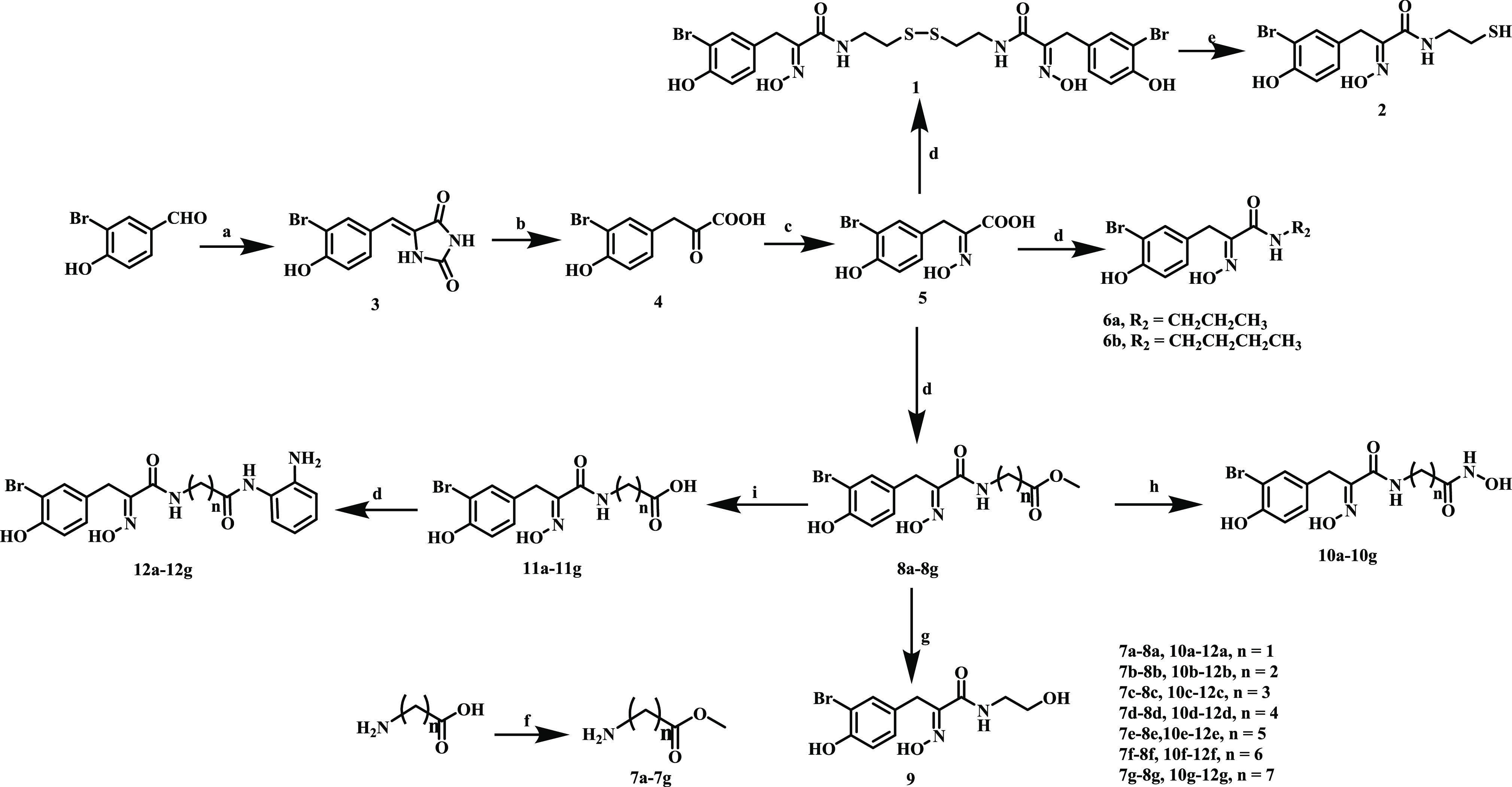
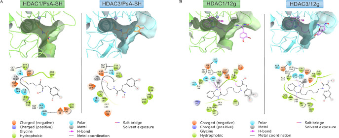
Psammaplin A (PsA) is a bromotyrosine disulfide dimer with histone deacetylase (HDAC) inhibition and acts through reduced monomer PsA-SH. We studied the connection of HDAC inhibition, cell growth inhibition, and apoptosis induction of PsA-SH by modifying the -SH group with deletion (6a) or replacement with hydroxamic acid (10b) or benzamide (12g). PsA-SH inhibits HDAC1/2/3 and 6a loses the HDAC inhibition ability. 10b inhibits HDAC1/2/3/6 while 12g shows selective inhibition of HDAC3. PsA-SH and 10b, but neither 6a nor 12g, induce apoptosis in human leukemia HL-60 cells associated with increased acetylation of Histone H3. PsA-SH and 10b inhibit growth of several solid tumor cell lines in vitro and Lewis lung cancer cell growth in vivo. PsA-SH is a simple scaffold for developing selective HDAC inhibitors and induces apoptosis through inhibiting HDAC1/2.


Conflict of interest statement

The authors declare no competing financial interest.

Figures

Figure 1
Figure 1
Chemical structures of Psammaplin A (PsA), Psammaplin A monomer (PsA-SH), and approved HDAC inhibitors.
Scheme 1
Scheme 1. Synthesis of PsA Monomer with a Different ZBG
Reagents and conditions: (a) Imidazolidine-2,4-dione, NaHCO3, H2O, 120 °C, 10 h, 83–92%; (b) NaOH, N2, H2O, 95 °C, 12 h, 65–73%; (c) NaOH, NaHCO3, HONH2·HCl, H2O, r.t., 12 h, 70–84%; (d) HOBt, EDCI, DIEA, DMF, 10 h, 30–47%; (e) DTT, KOH, MeOH, r.t., 30 min; (f) SOCl2, MeOH, Et2O, r.t., 3 h, 74–83%; (g) LiAlH4, THF, r.t., 1 h; (h) HONH2·H2O, NaOH, MeOH, r.t., 30 min, 73–88%; (i) NaOH, MeOH, 60 °C, 30 min, 90–95%.
Figure 2
Figure 2
FACS and Western blot analyses of apoptosis induction by PsA and PsA-SH in HL-60 cells. (A) HL-60 cells were treated with PsA and PsA-SH at the indicated concentrations for 24 h, and apoptotic cells were determined by FACS after staining with PI/Annexin-V. (B) Western blot analysis of the protein levels of Ac-H3, H3, Ac-α-tubulin, α-tubulin, PARP, caspase-3, -8, -9, and β-actin of HL-60 cells treated with PsA and PsA-SH for 24 h.
Figure 3
Figure 3
Apoptosis and H3 acetylation levels of HL-60 cells treated with 6a, 9, and 11b. Apoptotic cells were determined by FACS after staining with PI/Annexin-V (A), and the acetylated levels of H3 and α-tubulin were measured with Western blot (B) in HL-60 cells treated with each compound at 20 μM for 24 h.
Figure 4
Figure 4
Apoptosis induction and protein regulation by 10b and Tubacin. Apoptotic cells were determined by FACS after staining with PI/Annexin-V (A), and the protein levels of Ac-H3, Ac-α-tubulin, PARP, caspase-3, -8, -9, and β-actin were determined by Western blotting (B) in HL-60 cells treated with 10b and Tubacin at the indicated concentrations for 24 h.
Figure 5
Figure 5
Apoptosis induction and protein regulation by 12e, 12f, and RGFP966 in HL-60 cells. Apoptotic cells were determined by FACS after staining with PI/Annexin-V (A), and the protein levels of Ac-H3, Ac-α-tubulin, PARP, caspase-3, -8, -9, and β-actin were determined by Western blotting (B) in HL-60 cells treated with 12e, 12f, and RGFP966 for 24 h.
Figure 6
Figure 6
Putative binding patterns of PsA-SH (A) or 12g (B) within HDAC1 (left panel, green) and HDAC3 (right panel, blue) pockets.

Similar articles

Cited by

References

    1. Smith B. C.; Denu J. M. Chemical mechanisms of histone lysine and arginine modifications. Biochim. Biophys. Acta, Gene Regul. Mech. 2009, 1789, 45–57. 10.1016/j.bbagrm.2008.06.005. - DOI - PMC - PubMed
    1. Garmpis N.; Damaskos C.; Garmpi A.; Dimitroulis D.; Spartalis E.; Margonis G. A.; Schizas D.; Deskou I.; Doula C.; Magkouti E.; Andreatos N.; Antoniou E. A.; Nonni A.; Kontzoglou K.; Mantas D. Targeting Histone Deacetylases in Malignant Melanoma: A Future Therapeutic Agent or Just Great Expectations?. Anticancer research 2017, 37, 5355–5362. - PubMed
    1. Bezecny P. Histone deacetylase inhibitors in glioblastoma: pre-clinical and clinical experience. Medical oncology (Northwood, London, England) 2014, 31, 985.10.1007/s12032-014-0985-5. - DOI - PubMed
    1. Taunton J.; Hassig C. A.; Schreiber S. L. A mammalian histone deacetylase related to the yeast transcriptional regulator Rpd3p. Science (Washington, DC, U. S.) 1996, 272, 408–11. 10.1126/science.272.5260.408. - DOI - PubMed
    1. Xu W. S.; Parmigiani R. B.; Marks P. A. Histone deacetylase inhibitors: molecular mechanisms of action. Oncogene 2007, 26, 5541–52. 10.1038/sj.onc.1210620. - DOI - PubMed

LinkOut - more resources