. 2020 Dec 4;12(1):74-81.
doi: 10.1021/acsmedchemlett.0c00440. eCollection 2021 Jan 14.

Sweet and Blind Spots in E3 Ligase Ligand Space Revealed by a Thermophoresis-Based Assay

Affiliations

Sweet and Blind Spots in E3 Ligase Ligand Space Revealed by a Thermophoresis-Based Assay

Samuel Maiwald et al. ACS Med Chem Lett. .

Erratum in

Abstract

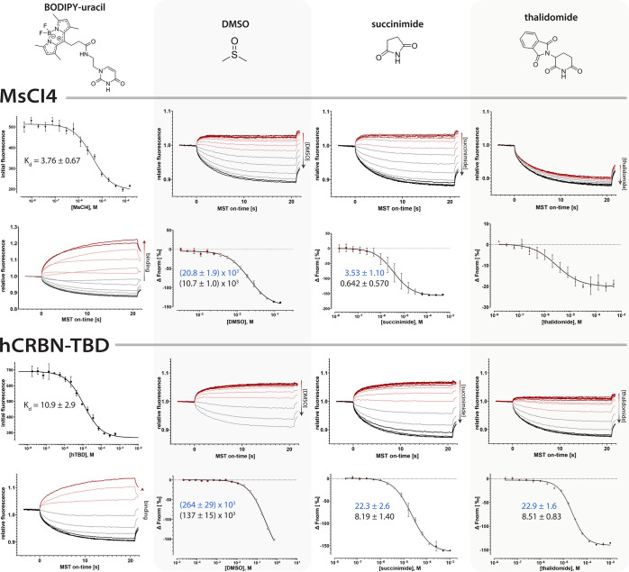
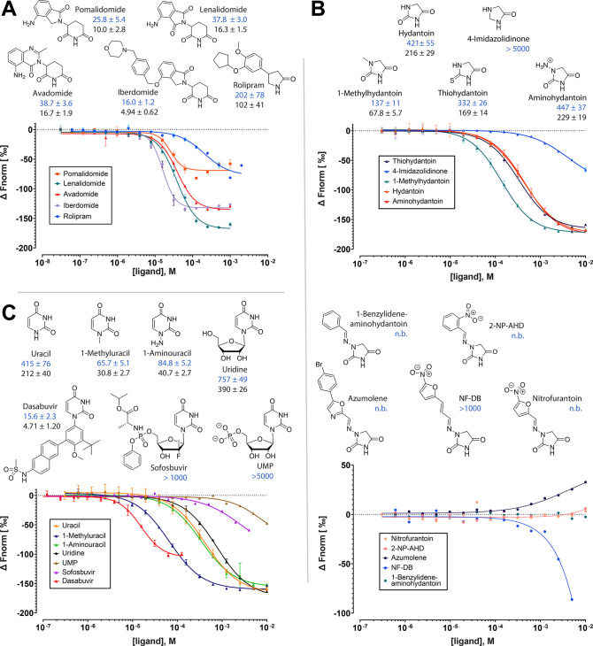
Repurposing E3 ubiquitin ligases for targeted protein degradation via customized molecular glues or proteolysis-targeting chimeras (PROTACs) is an increasingly important therapeutic modality. Currently, a major limitation in the design of suitable molecular glues and PROTACs is our fragmentary understanding of E3 ligases and their ligand space. We here describe a quantitative assay for the discovery and characterization of E3 ligase ligands that is based on the thermophoretic behavior of a custom reporter ligand. Thereby, it is orthogonal to commonly employed fluorescence-based assays and less affected by the optical properties of test compounds. It can be employed for the high-throughput screening of compound libraries for a given ligase but also for hit validation, which we demonstrate with the identification of unexpected well-binders and non-binders, yielding new insights into the ligand space of cereblon (CRBN).


Conflict of interest statement

The authors declare no competing financial interest.

Figures

Figure 1
Figure 1
Overall structure and ligand binding modes for E3 ligase substrate receptors VHL and CRBN. (A) Structure of VHL-Elongin B-Elongin C (VBC) complex with bound HIF1-α peptide (PDB: 1LM8). (B) Illustrative surface representation of the VHL binding groove with bound 11mer reporter, based on the same structure, highlighting the position of the fluorophore and hydroxyproline (Hyp). (C) Structure of the bacterial CRBN homologue MsCI4 bound to thalidomide (PDB: 4V2Y). (D) Illustrative surface representation of MsCI4 bound to BODIPY-uracil reporter. (E) Proposed clash of nitrofurantoin in the binding pocket of CRBN. Nitrofurantoin (green) was aligned with a structure of hydantoin in complex with MsCI4 (PDB: 5OH7) and shown in comparison to thalidomide (red). The protruding moiety of nitrofurantoin comes in close proximity to W99 of MsCI4. (F) Depiction of the pharmacophore-based nomenclature for 5- and 6-membered rings. R refers to the protruding moiety with the canonical exit vector.
Figure 2
Figure 2
MST traces and dose–response curves for Kd determination of short (FAM-11mer) and long (FAM-19mer) reporter peptide to VHL, and competition experiments with the short (11mer) and long (19mer) peptides. IC50 and derived Ki values are shown in blue and black, respectively, together with their confidence intervals. All values are in nM. FAM, fluorescein amidite; Hyp, hydroxyproline.
Figure 3
Figure 3
MST traces and affinities of the BODIPY-uracil reporter and reference compounds to the bacterial MsCI4 and the thalidomide binding domain of human CRBN (hTBD). The affinity of BODIPY-uracil was determined via initial fluorescence measurements. The MST traces show that binding or unbinding of the reporter yields an inversion of the thermophoretic behavior. The affinities of DMSO, succinimide (without DMSO), and thalidomide (with 0.5% DMSO) were determined via out-competition of the reporter in MST measurements, for which the MST traces and derived dose–response curves are shown. IC50 and derived Ki values are shown in blue and black, respectively, together with their confidence intervals. All values are in μM.
Figure 4
Figure 4
Determination of Z′ factor for hTBD. MST traces of solvent (0.5% DMSO) and positive control thalidomide (top panel) and scatter plot and resulting Z′ values at on-time 5s and on-time 20s (bottom panel). Means of positive and negative controls are shown by solid lines; dashed lines indicate 3-fold standard deviations.
Figure 5
Figure 5
Chemical structures, dose–response curves and affinity values for (A) IMiDs and rolipram, (B) small hydantoins and hydantoins branched via hydrazo groups, and (C) uracils to hTBD. IC50 and derived Ki values are shown in blue and black, respectively, together with their confidence intervals. All values are in μM. n.b., no binding; 2-NP-AHD, 1-(2-nitrobenzylideneamino)hydantoin; NF-DB, 1-(3-(5-nitrofuran-2-yl)allylidene)amino)hydantoin.

Similar articles

Cited by

References

    1. Lai A. C.; Crews C. M. Induced protein degradation: an emerging drug discovery paradigm. Nat. Rev. Drug Discovery 2017, 16 (2), 101–114. 10.1038/nrd.2016.211. - DOI - PMC - PubMed
    1. Chamberlain P. P.; Hamann L. G. Development of targeted protein degradation therapeutics. Nat. Chem. Biol. 2019, 15 (10), 937–944. 10.1038/s41589-019-0362-y. - DOI - PubMed
    1. Schapira M.; Calabrese M. F.; Bullock A. N.; Crews C. M. Targeted protein degradation: expanding the toolbox. Nat. Rev. Drug Discovery 2019, 18 (12), 949–963. 10.1038/s41573-019-0047-y. - DOI - PubMed
    1. Steinebach C.; Kehm H.; Lindner S.; Vu L. P.; Kopff S.; Marmol A. L.; Weiler C.; Wagner K. G.; Reichenzeller M.; Kronke J.; Gutschow M. PROTAC-mediated crosstalk between E3 ligases. Chem. Commun. 2019, 55 (12), 1821–1824. 10.1039/C8CC09541H. - DOI - PubMed
    1. Girardini M.; Maniaci C.; Hughes S. J.; Testa A.; Ciulli A. Cereblon versus VHL: Hijacking E3 ligases against each other using PROTACs. Bioorg. Med. Chem. 2019, 27 (12), 2466–2479. 10.1016/j.bmc.2019.02.048. - DOI - PMC - PubMed