. 2020 Dec 25;12(1):107-114.
doi: 10.1021/acsmedchemlett.0c00556. eCollection 2021 Jan 14.

Solution Conformations Shed Light on PROTAC Cell Permeability

Affiliations

Solution Conformations Shed Light on PROTAC Cell Permeability

Yoseph Atilaw et al. ACS Med Chem Lett. .

Abstract

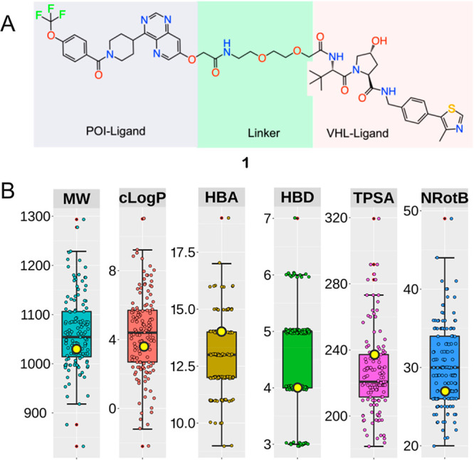
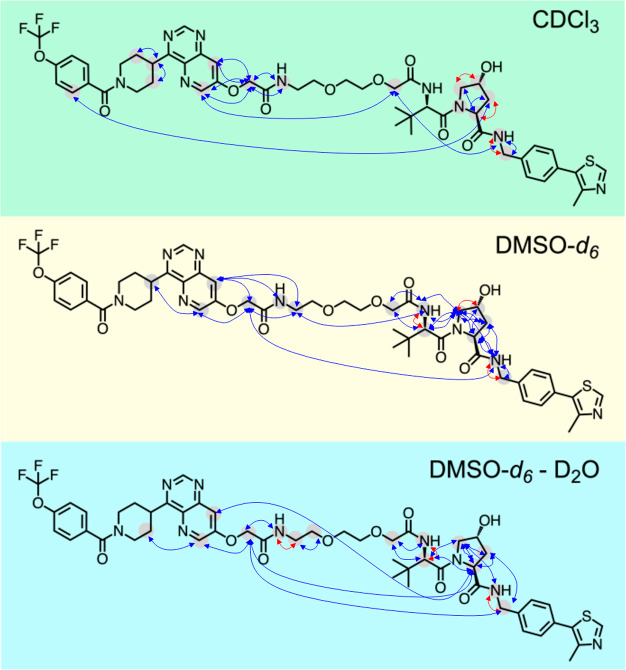
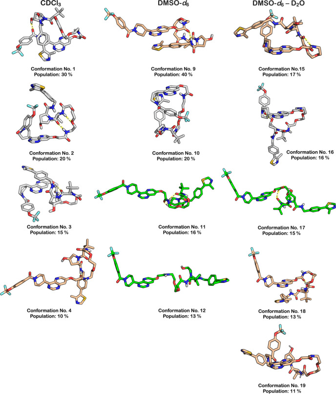
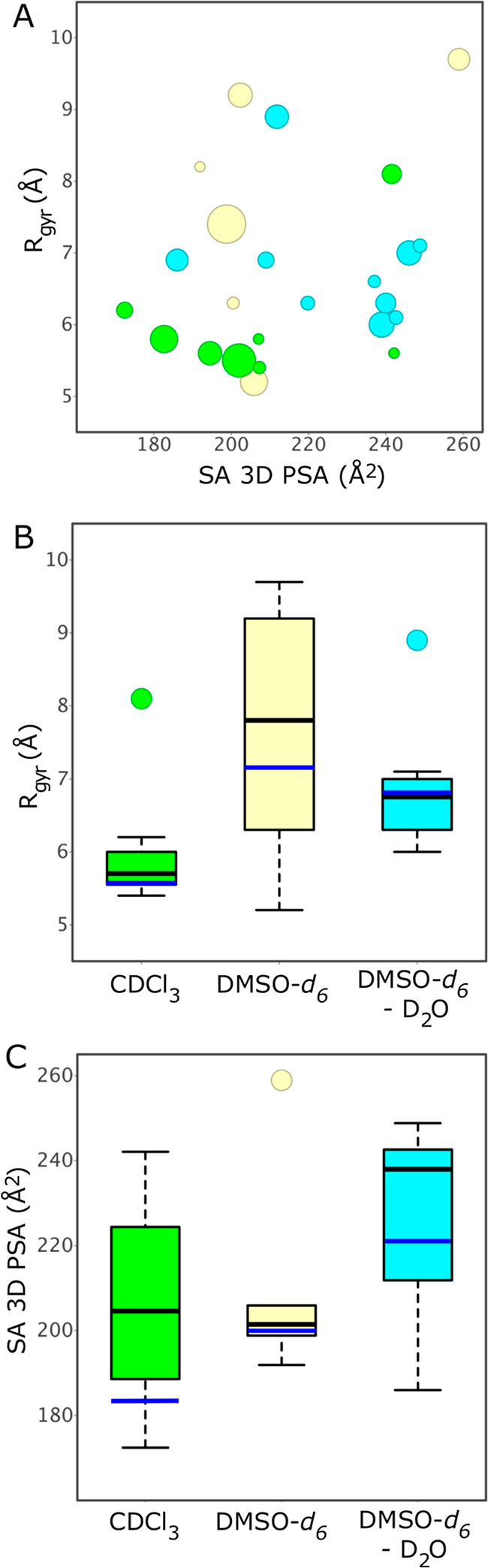
Proteolysis targeting chimeras (PROTACs) induce intracellular degradation of target proteins. Their bifunctional structure puts degraders in a chemical space where ADME properties often complicate drug discovery. Herein we provide the first structural insight into PROTAC cell permeability obtained by NMR studies of a VHL-based PROTAC (1), which is cell permeable despite having a high molecular weight and polarity and a large number of rotatable bonds. We found that 1 populates elongated and polar conformations in solutions that mimic extra- and intracellular compartments. Conformations were folded and had a smaller polar surface area in chloroform, mimicking a cell membrane interior. Formation of intramolecular and nonclassical hydrogen bonds, π-π interactions, and shielding of amide groups from solvent all facilitate cell permeability by minimization of size and polarity. We conclude that molecular chameleonicity appears to be of major importance for 1 to enter into target cells.


Conflict of interest statement

The authors declare the following competing financial interest(s): D.G. is an employee of Nuvisan Innovation Campus Berlin GmbH. A.G and D.M. are employees of Bayer AG.

Figures

Figure 1
Figure 1
(A) Structure of PROTAC 1. The ligand for the protein target of interest (POI), ERK5, the linker, and the ligand for binding to the E3 ligase (VHL) are indicated by gray, green, and pink shading, respectively. (B) Molecular weight (MW, Da), lipophilicity (cLogP), hydrogen bond acceptors and donors (HBA and HBD), topological polar surface area (TPSA, Å2), and number of rotatable bonds (NRotB) calculated for PROTAC 1 and the subset of 135 VHL PROTACs that have PEG-based linkers reported by Maple et al. The values of the six descriptors calculated for 1 are shown as yellow circles (MW = 1034 Da, cLogP = 3.55, HBA = 14, HBD = 4, TPSA = 237 Å2, NRotB = 27). Box plots show the 50th percentiles as horizontal bars, the 25th and 75th percentiles as boxes, the 25th percentile minus 1.5× the interquartile range, and the 75th percentile plus 1.5× the interquartile range as whiskers for the subset of PROTACs having PEG-based linkers. Outliers are shown both as red dots and as circles in the color of the appropriate descriptor.
Scheme 1
Scheme 1. Synthesis of PROTAC 1
Reagents and conditions: (a) Ethyl bromoacetate, K2CO3, THF, 100 °C, 8 h; (b) aq. NaOH, EtOH, rt, overnight, 38% from 2; (c) HATU, N,N-diisopropylethylamine, DMF, rt, overnight, 48%.
Figure 2
Figure 2
Overview of experimentally determined proton–proton distances and dihedral angles that were used to determine the solution conformational ensembles of 1. Blue arrows indicate proton–proton distances, while dihedral angles are indicated by red arrows.
Figure 3
Figure 3
Structures of the major conformations (population ≥ 10%) in the ensembles adopted by 1 in chloroform, DMSO, and DMSO–water (10:1). Conformations that are predominantly linear are in green, those that are folded with one turn are in tan, while folded conformations with two turns are in gray. The number and population in percent is given below each conformation. Intramolecular hydrogen bonds are indicated with black dotted lines on a yellow background.
Figure 4
Figure 4
(A) Radius of gyration (Rgyr) versus solvent accessible 3D polar surface area (SA 3D PSA) for all solution conformations populated by 1. The area of each circle is proportional to the population of the corresponding conformation (in %). Conformations in CDCl3 are in green, those in DMSO-d6 are in yellow, while those in DMSO-d6–D2O are in cyan. (B) Radius of gyration (Rgyr) and (C) solvent accessible 3D polar surface area (SA 3D PSA) for the conformations populated by 1 in CDCl3, DMSO-d6, and DMSO-d6–D2O. Box plots show the 50th percentiles as black horizontal bars, the 25th and 75th percentiles as boxes, the 25th percentile minus 1.5× the interquartile range, and the 75th percentile plus 1.5× the interquartile range as whiskers, and outliers as colored circles. Population weighted mean values are shown as blue horizontal bars.
Figure 5
Figure 5
Structures of conformations 1–3 that are populated in chloroform. The population in percent, the number of intramolecular hydrogen bonds (IMHB), the solvent accessible 3D PSA and the radius of gyration (Rgyr) are given below each conformation. IMHBs are indicated with dashed black lines on a yellow background.

Similar articles

Cited by

References

    1. Lai A. C.; Crews C. M. Induced protein degradation: an emerging drug discovery paradigm. Nat. Rev. Drug Discov. 2017, 16, 101–114. 10.1038/nrd.2016.211. - DOI - PMC - PubMed
    1. Schapira M.; Calabrese M. F.; Bullock A. N.; Crews C. M. Targeted protein degradation: expanding the toolbox. Nat. Rev. Drug Discov. 2019, 18, 949–963. 10.1038/s41573-019-0047-y. - DOI - PubMed
    1. Bondeson D. P.; Mares A.; Smith I. E. D.; Ko E.; Campos S.; Miah A. H.; Mulholland K. E.; Routly N.; Buckley D. L.; Gustafson J. L.; Zinn N.; Grandi P.; Shimamura S.; Bergamini G.; Faelth-Savitski M.; Bantscheff M.; Cox C.; Gordon D. A.; Willard R. R.; Flanagan J. J.; Casillas L. N.; Votta B. J.; den Besten W.; Famm K.; Kruidenier L.; Carter P. S.; Harling J. D.; Churcher I.; Crews C. M. Catalytic in vivo protein knockdown by small-molecule PROTACs. Nat. Chem. Biol. 2015, 11, 611–617. 10.1038/nchembio.1858. - DOI - PMC - PubMed
    1. Testa A.; Hughes S. J.; Lucas X.; Wright J. E.; Ciulli A. Structure-based design of a macrocyclic PROTAC. Angew. Chem., Int. Ed. 2020, 59, 1727–1734. 10.1002/anie.201914396. - DOI - PMC - PubMed
    1. Ermondi G.; Vallaro M.; Caron G. Degraders early developability assessment: face-to-face with molecular properties. Drug Discov. Today 2020, 25, 1585.10.1016/j.drudis.2020.06.015. - DOI - PubMed