. 2020 Dec 29:19:384-400.
doi: 10.1016/j.csbj.2020.12.017. eCollection 2021.

Microbial risk assessment of Nocardia cyriacigeorgica in polluted environments, case of urban rainfall water

Affiliations

Microbial risk assessment of Nocardia cyriacigeorgica in polluted environments, case of urban rainfall water

Florian Vautrin et al. Comput Struct Biotechnol J. .

Abstract

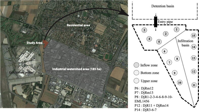
Urban Infiltration Basins (UIBs) are used to manage urban runoff transfers and feed aquifers. These UIBs can accumulate urban pollutants and favor the growth of potentially pathogenic biological agents as Nocardia.

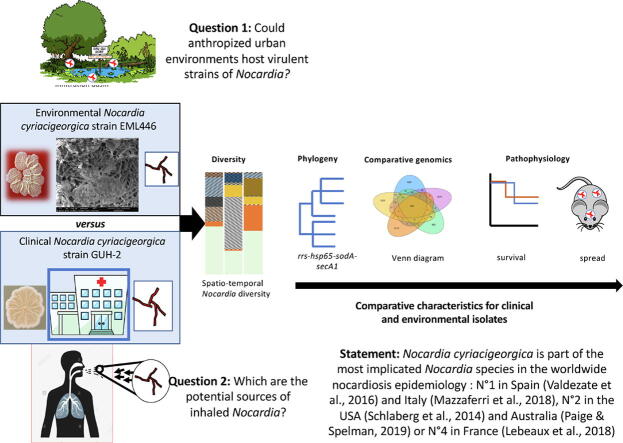
Objectives: To assess the spatio-temporal dynamics of pathogenic Nocardia in UIBs and to stablish phylogenetic relationships between clinical and UIB N. cyriacigeorgica strains. To assess pathogenicity associated with environmental N. cyriacigeorgica using an animal model, and to identify genetic elements that may be associated to its virulence.

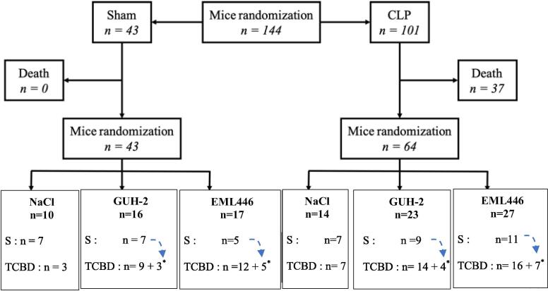
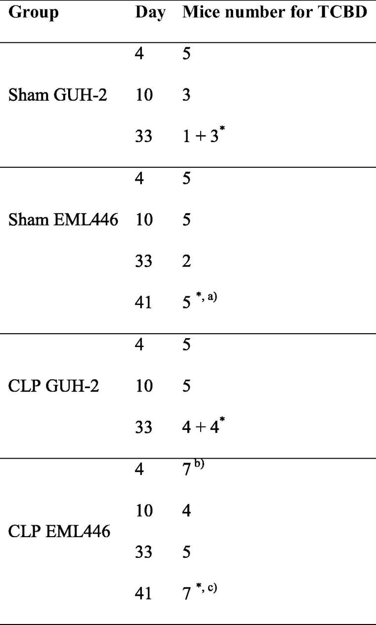
Methods: A well-characterized UIB in terms of chemical pollutants from Lyon area was used in this study during a whole year. Cultural and Next-Generation-Sequencing methods were used for Nocardia detection and typing. Clinical and environmental isolates phylogenetic relationships and virulences were compared with Multilocus-Sequence-Analysis study together with a murine model.

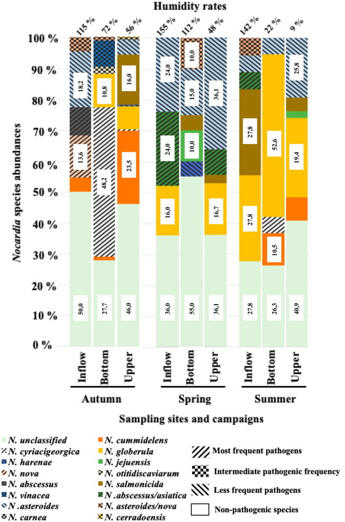
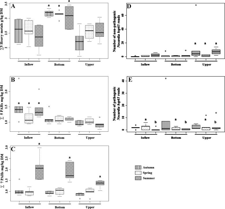
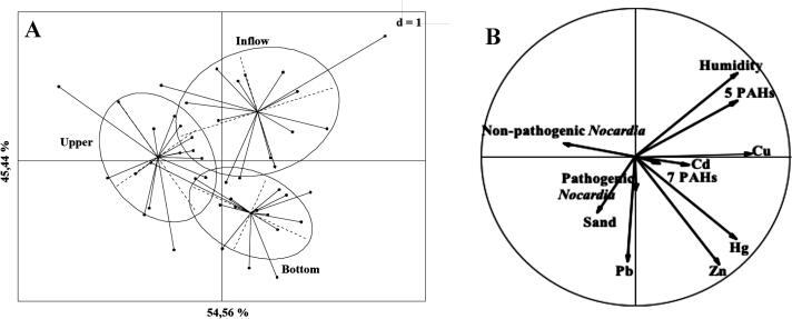
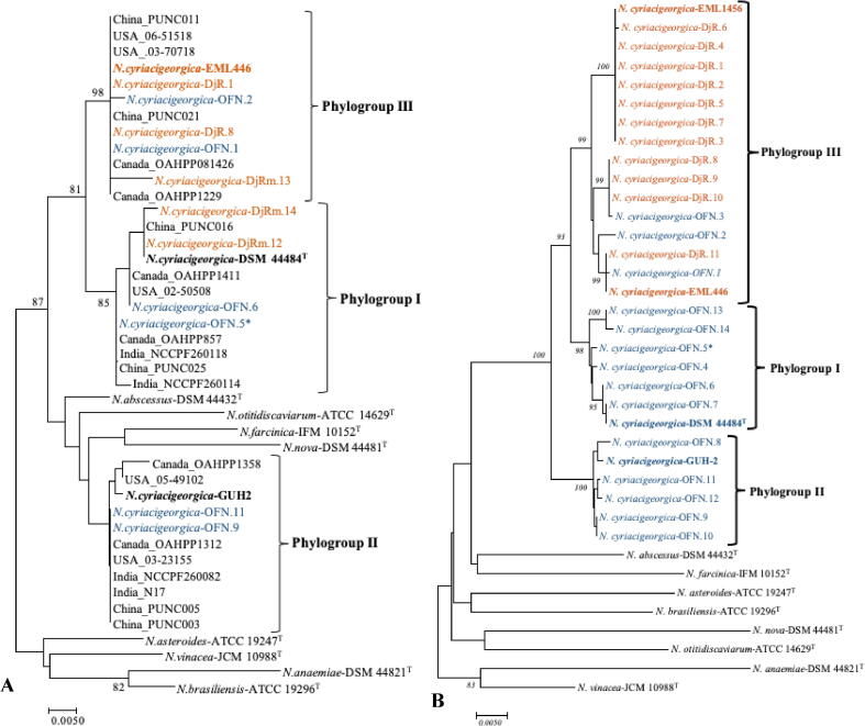
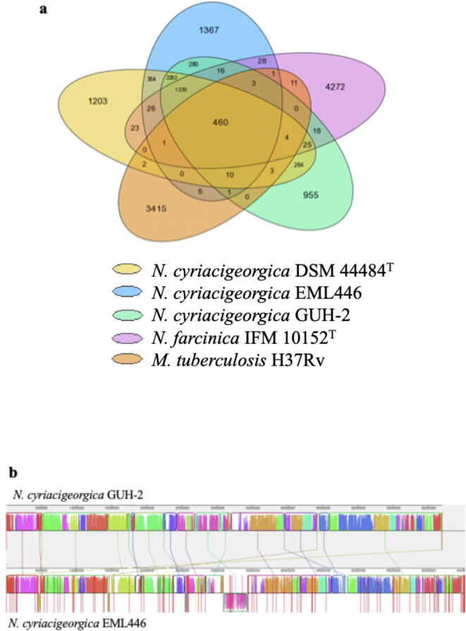
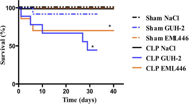
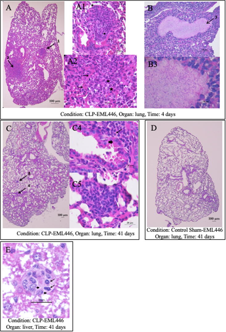
Results: In autumn, N. cyriacigeorgica and N. nova were the pathogenic most prevalent species in the UIB. The complex N. abscessus/asiatica was also detected together with some other non-pathogenic species. The presence of pathogenic Nocardia was positively correlated to metallic trace elements. Up to 1.0 × 10³ CFU/g sediment of N. cyriacigeorgica and 6 OTUs splited in two different phylogroups were retrieved and were close to clinical strains. The EML446 tested UIB isolate showed significant infectivity in mice with pulmonary damages similar to clinical clone (GUH-2).

Conclusion: Hsp65 marker-based metabarcoding approach allowed detecting N. cyriacigeogica as the most abundant Nocardia pathogenic species in a UIB. Metal trace elements-polluted environments can be reservoirs of pathogenic Nocardia which may have a similar virulence to clinical strains.

Keywords: Environment; Hsp65 metabarcoding; Murine model of transient immunoparalysis; Nocardia; Opportunistic pathogen; Urban pollution.


Conflict of interest statement

The authors declare that they have no known competing financial interests or personal relationships that could have appeared to influence the work reported in this paper.

Figures

None
Graphical abstract
Fig. 1
Fig. 1
Aerial image of the Django-Reinhardt infiltration basin (DRIB) and position of the sampling points in the DRIB and placement of the three different sampling areas (inflow zone, bottom zone and upper zone). Px: DRIB sample point in which N. cyriacigeorgica was isolated and respective reference code.
Fig. 2A
Fig. 2A
Study design describing the experiment made with the immunosuppressed CLP (cecal ligature and puncture) (30%) murine model to compare the virulence of the SIS (stormwater infiltration system) and clinical N. cyriacigeorgica representative strains. Sham = mice that underwent laparotomy with exposition of the cecum but without CLP. CLP = intestinal tract externalization and puncture performed as described by Restagno et al. . S = Survival experiment. TCBD = Time Course Bacterial Detection experiment. n = number of individuals in each group. * = surviving mice at the end of the ‘S’ experiment retrieved for ‘TCBD’ one at D33 (GUH-2) or D41 (EML446).
Fig. 2B
Fig. 2B
Mice distribution for the Time Course Bacterial Detection experiment (TCBD). * = Surviving mice from the end of the Survival experiment retrieved at the end of the TCBD one. a) lungs of two mice will be reserved for histological analysis, b) lungs of one mouse will be reserved for histological analysis. c) lungs of three mice and brains of two mice will be reserved for histological analysis.
Fig. 3
Fig. 3
DNA metabarcoding analysis of Nocardia genetic diversity among SIS (stormwater infiltration system) sediment samples using the hsp65 target. A total of 7,128 sequences per sample was analyzed. The relative proportion of the detected Nocardia species is presented in %. Species representing>1% over the total number of reads are indicated and only values > 10% are indicated. The classification of most/intermediate/less frequent pathogens was based on the most clinically relevant species in France epidemiology according to Lebeaux et al. .
Fig. 4
Fig. 4
Boxplots explicative of the between-class analysis (BCA). A. 5 trace element metals: Cd (Cadmium); Cu (Copper); Hg (Mercury); Pb (Lead) and Zn (Zinc). P-value a = 0.0195. B. 5 PAHs: fluoranthene, pyrene, phenanthrene, benzo(a)anthracene, benzo(a)pyrene. P-value a = 0.0157. C. 7 PAHs (polycyclic aromatic hydrocarbons): naphthalene, acenaphthene, fluorene, benzo(b)fluoranthene, dibenzo(a,h)anthracene, benzo(ghi)perylene, indeno(1,2,3-cd)pyrene. P-value a = 6.97x10⁻⁶. D. Non-pathogenic Nocardia species: N. cummidelens, N. globerula, N. harenae, N. iowensis, N. jejuensis, N. pseudovaccinii, N. salmonicida, N. soli. P-value a = 0.0294. E. Pathogenic Nocardia species: N. abscessus, N. abscessus/asiatica, N. anaemiae, N. asteroides, N. brasiliensis, N. carnea, N. cerradoensis, N. cyriacigeorgica, N. ninae, N. nova, N. otitidiscaviarum, N. shimofusensis, N. sienata, N. vinaceae. P-values a = 0.04055 and b = 0.02963.
Fig. 5
Fig. 5
A. Between-Class Analysis (BCA) performed on physical–chemical parameters and Nocardia relative abundance from sediments of the Django-Reinhardt infiltration basin (DRIB). B. Explicative factors of the BCA. 7 PAHs (polycyclic aromatic hydrocarbons): naphthalene, acenaphthene, fluorene, benzo(b)fluoranthene, dibenzo(a,h)anthracene, benzo(ghi)perylene, indeno(1,2,3-cd)pyrene ; 5 PAHs: fluoranthene, pyrene, phenanthrene, benzo(a)anthracene, benzo(a)pyrene; Cd (Cadmium); Cu (Copper); Hg (Mercury); Pb (Lead) and Zn (Zinc). Pathogenic Nocardia species: N. abscessus, N. abscessus/asiatica, N. anaemiae, N. asteroides, N. brasiliensis, N. carnea, N. cerradoensis, N. cyriacigeorgica, N. ninae, N. nova, N. otitidiscaviarum, N. shimofusensis, N. sienata, N. vinaceae ; non-pathogenic Nocardia species: N. cummidelens, N. globerula, N. harenae, N. iowensis, N. jejuensis, N. pseudovaccinii, N. salmonicida, N. soli.
Fig. 6
Fig. 6
Correlogram of the physical–chemical parameters and Nocardia pathogenic and non-pathogenic species relative abundance. 7 PAHs (polycyclic aromatic hydrocarbons): naphthalene, acenaphthene, fluorene, benzo(b)fluoranthene, dibenzo(a,h)anthracene, benzo(ghi)perylene, indeno(1,2,3-cd)pyrene ; 5 PAHs: fluoranthene, pyrene, phenanthrene, benzo(a)anthracene, benzo(a)pyrene; trace element metals: Cd (Cadmium); Cu (Copper); Hg (Mercury); Pb (Lead) and Zn (Zinc).
Fig. 7
Fig. 7
Molecular phylogeny of Nocardia cyriacigeorgica. A) Unrooted phylogenetic tree based on the hsp65 sequences (401 bp) from USA , Canada , India and China isolates, and representative N. cyriacigeorgica sequences from the metabarcoding analysis, and the clinical and SIS strains reported in this study. OFN.5* corresponds to a patient from Lyon (France). DjR.1, DjR.8 and EML446 corresponding hsp65 sequences were used to represent the environmental cluster. B) Unrooted phylogenetic tree based on the rrs-hsp65-sodA-secA1 concatenated sequences (1,845 bp). The maximum likelihood tree was constructed using MEGA software (version 7.0.16) after having aligned the sequences with ClustalW. The bootstrap values were calculated from 1,000 replicates, and those higher than 80% are given at the corresponding nodes. Clinical strains are colored in blue, and environmental strains are colored in orange. (For interpretation of the references to color in this figure legend, the reader is referred to the web version of this article.)
Fig. 8
Fig. 8
Comparison of Nocardia cyriacigeorgica EML446 genome with other actinomycetal reference genomes. (a) Venn Diagram representing the number of shared CDSs between whole genomes of N. cyriacigeorgica DSM 44484T, N. cyriacigeorgica EML446, N. cyriacigeorgica GUH-2, N. farcinica IFM 10152T and Mycobacterium tuberculosis H37Rv. (b) MAUVE comparison between EML446 and GUH-2 genomes showing the position of RGPs (regions of genomic plasticity).
Fig. 9
Fig. 9
Survival rate after N. cyriacigeorgica cells instillations of Sham and CLP mice. Five days post-CLP (i.e. D0), mice were challenged with an intratracheal administration of Nocardia GUH-2 or EML446 at 1.0 × 106 CFU/mouse. Either NaCl (physiological saline solution) (Sham n = 7, CLP n = 7), GUH-2 (Sham n = 7, CLP n = 9) or EML446 (Sham n = 5, CLP n = 11) were instilled. Results are expressed as Kaplan-Meier survival curves. * p-value < 0.05 was considered statistically significant compared to the respective control groups.
Fig. 10
Fig. 10
Histological lung and liver sections of CLP-EML446 mouse at days 4 and 41 after intratracheal instillation. A, B: CLP-EML446 mouse lung at day 4. Light micrographs of mouse lung section with evidence of granulomatous process characterized by both inflammatory response (A) and necrosis usually present in nocardiosis (B). A: Two early pyogranulomas disseminated in the pulmonary parenchyma (arrows). Original magnification: obj. 4×, lungs, postcaval lobe. A1: enlargement of the pyogranulomas (arrow 1). Multiple degenerated neutrophilic polymorphonuclear cells into a lung alveolus lumen admixed with few macrophages. Some inflammatory cells overflow in the lumen of a bronchus. Thin arrow: bronchus epithelium. Large arrow: neutrophilic polymorphonuclear cells. Star: macrophages. Original magnification: obj. 40 X. A2: enlargement of the pyogranulomas (arrow 2). Multiple degenerated neutrophilic polymorphonuclear cells into a lung alveolus lumen admixed with few macrophages. Thin arrows: alveolar wall. Large arrow: neutrophilic polymorphonuclear cells. Star: macrophages. Original magnification: obj. 40 X. B: The lungs showed cavitary lesions constituted by central necrotic material surrounded by some polymorphonuclear and numerous macrophages. Macrophage alveolitis and interstitial lymphocytic infiltration are also observed. Light micrographs; hematoxylin–eosin staining; original magnification: obj. 40 X. B3: High power magnification showing histologic sections of the lesion characterized by caseous necrotic central area with filamentous bacteria inside. Light micrographs; hematoxylin–eosin staining; original magnification: obj. 100 X. C: CLP-EML446 mouse lung at day 41: C: At day 41, some lymphocyte aggregates are present in the interstitium (arrows). Original magnification: obj. 4×, lungs, postcaval lobe. C4: enlargement of the aggregate (arrow 4). Some small lymphocytes (thin arrow) infiltrate the interstitium beneath the bronchial epithelium (large arrow). Original magnification: obj. 100 X. C5: enlargement of the aggregate (arrow 5). Some small lymphocytes infiltrate the interstitium of the interalveolar wall. Original magnification: obj. 100 X. D: Sham EML446 mouse lung at day 41. no lesions. Original magnification obj. 4×, lungs, postcaval lobe. E: CLP-EML446 mouse liver at day 41. Mature granuloma disseminated at random into liver lobules. Thin arrows: lymphocytes. Large arrows: macrophages. Original magnification obj. 100×.

Similar articles

Cited by

References

    1. Brown-Elliott B.A., Brown J.M., Conville P.S., Wallace R.J., Jr Clinical and Laboratory Features of the Nocardia spp. Based on Current Molecular Taxonomy. CMR. 2006;19(2):259–282. doi: 10.1128/CMR.19.2.259-282.2006. - DOI - PMC - PubMed
    1. Rodriguez-Nava V., Durupt S., Chyderiotis S., Freydière A.-M., Karsenty J., de Montclos M., Reix P., Durieu I., Nove-Josserand R., Chiron R., Bremont F., Têtu L., Murris M., Terru D., Godreuil S., Bergeron E., Freney J., Boiron P., Vandenesch F., Marchandin H., Segonds C., Doléans-Jordheim A. A French multicentric study and review of pulmonary Nocardia spp. in cystic fibrosis patients. Med Microbiol Immunol. 2015;204(4):493–504. doi: 10.1007/s00430-014-0360-3. - DOI - PubMed
    1. Heise E.R. Diseases associated with immunosuppression. Environ Health Perspect. 1982;43:9–19. doi: 10.1289/ehp.82439. - DOI - PMC - PubMed
    1. Luo Q., Hiessl S., Poehlein A., Daniel R., Steinbüchel A., Loeffler F.E. Insights into the microbial degradation of rubber and gutta-percha by analysis of the complete genome of Nocardia nova SH22a. Appl Environ Microbiol. 2014;80(13):3895–3907. doi: 10.1128/AEM.00473-14. - DOI - PMC - PubMed
    1. Luo Q., Hiessl S., Steinbüchel A. Functional diversity of Nocardia in metabolism: metabolism of nocardia. Environ Microbiol. 2014;16(1):29–48. doi: 10.1111/1462-2920.12221. - DOI - PubMed

LinkOut - more resources