. 2020 Dec;23(12):1527-1537.
doi: 10.22038/ijbms.2020.43303.10168.

Design, synthesis, and biological evaluation of novel 5,6,7-trimethoxy quinolines as potential anticancer agents and tubulin polymerization inhibitors

Affiliations

Design, synthesis, and biological evaluation of novel 5,6,7-trimethoxy quinolines as potential anticancer agents and tubulin polymerization inhibitors

Salimeh Mirzaei et al. Iran J Basic Med Sci. 2020 Dec.

Abstract

Objectives: Microtubules have key roles in essential cellular processes such as mitosis, cell motion, and intracellular organelle transport. Increasing interest has been given to tubulin binding compounds after the introduction of taxanes into clinical oncology. The object of this study was synthesis and biological evaluation of novel 5,6,7-trimethoxy quinolines as tubulin inhibitors.

Materials and methods: The cytotoxicity of the newly synthesized compounds was assessed against different human cancer cell lines including MCF-7, A2780, MCF-7/MX, A2780/RCIS, and normal cells. Compounds demonstrating the most antiproliferative activity, were chosen to examine their tubulin inhibition activity and their ability to arrest the cell cycle and induce apoptosis. Molecular docking studies and molecular dynamics simulation of compound 7e in the catalytic site of tubulin were performed.

Results: Most of the synthesized quinolines showed moderate to significant cytotoxic activity against human cancer cells. Compounds 7e and 7f, possessing N-(4-benzoyl phenyl) and N-(4-phenoxy phenyl), respectively, exhibited the most antiproliferative activity more potent than the other compounds and exhibited similar antiproliferative activity on both resistant and parental cancer cells.

Conclusion: Flow cytometry analysis of A2780, A2780/RCIS, MCF-7, and MCF-7/MX cancer cells treated with 7e and 7f exhibited that these compounds arrested the cell cycle (at the G2/M phase) and induced cellular apoptosis in A2780 cancer cells. These quinolines inhibited tubulin polymerization in a way resembling that of CA-4. Molecular dynamics simulation and molecular docking studies of compound 7e into the binding site of tubulin displayed the probable interactions of 7e with the binding site of tubulin.

Keywords: Anticancer activity; Apoptosis; Quinoline; Resistant cancer cells; Tubulin inhibitors.


Figures

Figure 1
Figure 1
Reported anticancer agents and novel trimethoxy quinolines as tubulin inhibitors
Figure 2
Figure 2
Effect of compounds 7e and 7f on in vitro tubulin polymerization
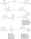
Scheme 1
Scheme 1
Reagents and conditions: (a) 120° C, 2 hr (b) POCl3, 120 oC, 2 hr (c) absolute ethanol, reflux, 4–12 hr, (d) para toluene sulfonamide, toluene, 140 °C, reflux
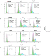
Figure 3
Figure 3
Flow cytometry analysis of compound 7f, in human cancer cell lines: (A) A2780, (B) A2780/RCIS, (C) MCF-7, (D) MCF-7/MX
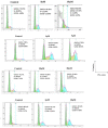
Figure 4.
Figure 4.
Flow cytometry analysis of compound 7e in human cancer cell lines, (A) A2780, (B) A2780/RCIS, (C) MCF-7, (D) MCF-7/MX
Figure 5
Figure 5
Effect of 7e on the apoptosis of A2780 cancer cells assessed using Annexin V/PI double staining test by flow cytometry. The percentages of cells in each stage of apoptosis were quantitated by flow cytometry
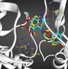
Figure 6
Figure 6
Compounds 7e in red, 7f in yellow, and 7g in blue docked in the active site of tubulin
Figure 7
Figure 7
RMSD between protein with ligand and without ligand
Figure 8
Figure 8
RMSD per residue of protein with ligand and without ligand during 80 nsec simulation
Figure 9
Figure 9
Rg of protein with ligand and without ligand
Figure 10
Figure 10
Number of hydrogen bonds between protein and ligand in time scale
Figure 11
Figure 11
Number of intramolecular hydrogen bonds of protein in the time scale
Figure 12
Figure 12
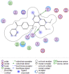
The 2D representation of the interaction between compound 7e in the crystal structure of tubulin (PDB ID: 4O2B) after 80 nsec molecular dynamic simulation using LigX in MOE

Similar articles

Cited by

References

    1. Kirschner M, Mitchison T. Beyond self-assembly: from microtubules to morphogenesis. Cell. 1986;45:329–342. - PubMed
    1. Mignot I, Pecqueur L, Dorléans A, Karuppasamy M, Ravelli RB, Dreier B, et al. Design and characterization of modular scaffolds for tubulin assembly. J Biol Chem. 2012;287:31085–31094. - PMC - PubMed
    1. Akhmanova A, Steinmetz MO. Control of microtubule organization and dynamics: two ends in the limelight. Nat Rev Mol Cell Biol. 2015;16:711–726. - PubMed
    1. Kadavath H, Hofele RV, Biernat J, Kumar S, Tepper K, Urlaub H, et al. Tau stabilizes microtubules by binding at the interface between tubulin heterodimers. Proc Natl Acad Sci. 2015;112:7501–7506. - PMC - PubMed
    1. Chandrasekaran G, Tátrai P, Gergely F. Hitting the brakes: targeting microtubule motors in cancer. Br J Cancer. 2015;113:693–698. - PMC - PubMed

LinkOut - more resources