. 2020 Dec;23(12):1590-1602.
doi: 10.22038/ijbms.2020.41550.11078.

N-acetylcysteine-loaded electrospun mats improve wound healing in mice and human fibroblast proliferation in vitro: a potential application of nanotechnology in wound care

Affiliations

N-acetylcysteine-loaded electrospun mats improve wound healing in mice and human fibroblast proliferation in vitro: a potential application of nanotechnology in wound care

Ramin Seyedian et al. Iran J Basic Med Sci. 2020 Dec.

Abstract

Objectives: N-acetylcysteine (NAC) has gained attention recently in dermatology as a unique anti-oxidant. In light of progress in nanotechnological methods, it was hypothesized that loading NAC onto nanofibers would positively affect skin wound healing. The objective of this study was to fabricate NAC-loaded electrospun mats and test their effect on wound healing in vivo and in vitro.

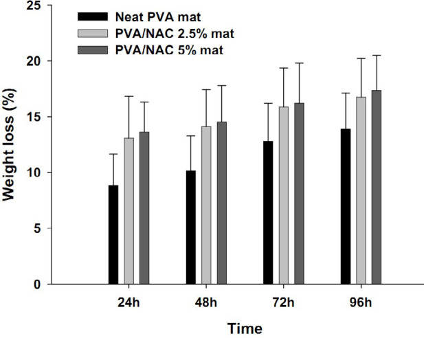
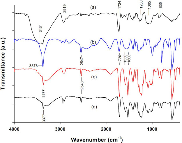
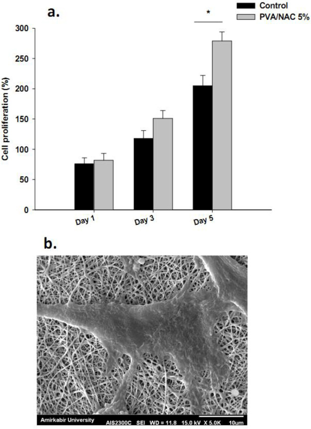
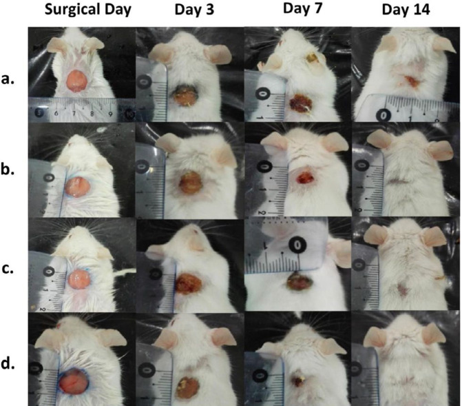
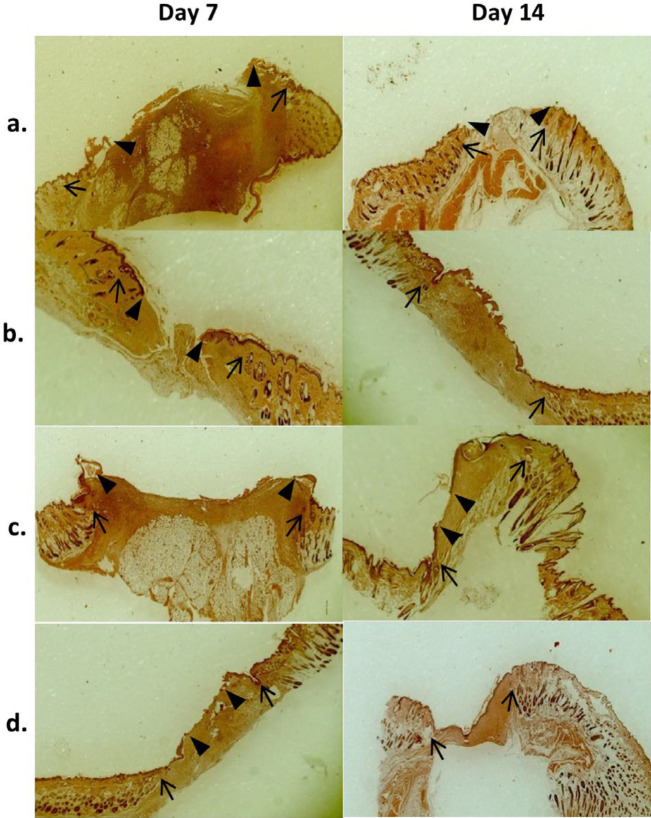
Materials and methods: Polyvinyl alcohol (PVA)-based mats loaded with NAC at three concentrations were electrospun and characterized in terms of physicochemical properties and drug release profile. Human fibroblast cells (in vitro) and mouse full-thickness skin wounds (in vivo) were treated with mats for 5 and 14 days, respectively. Wound area, tissue histopathology, fibroblast proliferation and cellular oxidative state were evaluated.

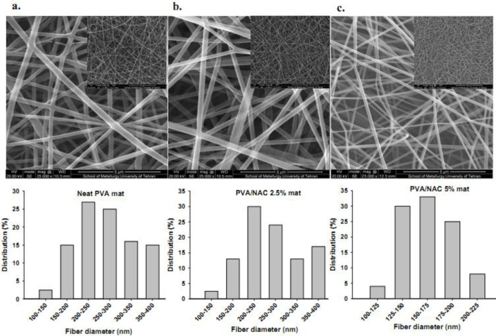
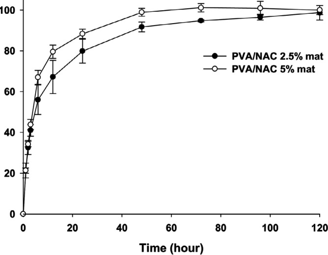
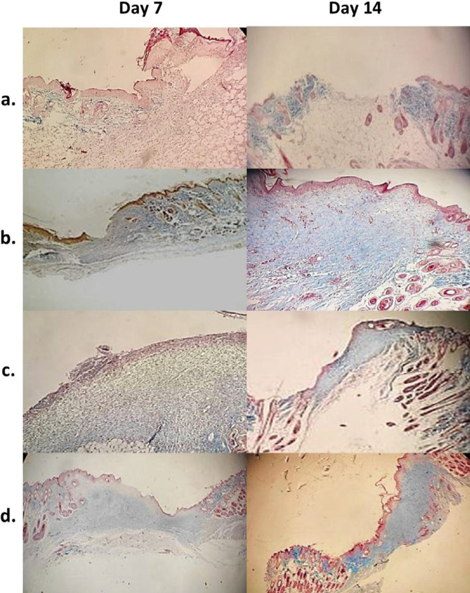
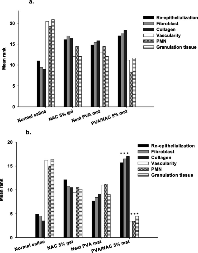
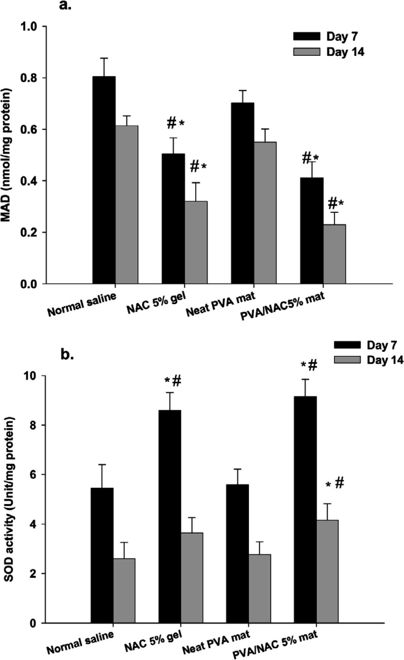
Results: Mats containing 5% PVA/NAC showed thinner fibers with suitable physicochemical properties and a sustained drug release profile. PVA/NAC (5%) mats enhanced fibroblast proliferation and attachment in vitro. The mats resulted in significant wound closure with high levels of re-epithelialization and collagen fiber synthesis on day 14 post-surgery in vivo. The mats also reduced granulation tissue and edematous stroma to a higher extent. These findings were accompanied by a significant decrease in tissue lipid peroxidation and higher superoxide dismutase activity, which may explain how NAC improved wound healing.

Conclusion: We propose an NAC-loaded nanofibrous mat that takes the advantage of a porous nanoscaffold structure to release NAC in a sustained manner. This mat may be a promising candidate for further clinical evaluation.

Keywords: Electrospun nanofiber; In vitro; Mouse; N-acetylcysteine (NAC); Oxidative stress; Wound.


Figures

Figure 1
Figure 1
Morphology and the corresponding size distribution of nanofibers in neat PVA, PVA/NAC 2.5% and PVA/NAC 5% mats. Scale bars represent 5 µm in the large SEM photos. Inset photos at lower magnifications are also provided for each mat type. NAC: N-acetylcysteine; PVA: Polyvinyl alcohol
Figure 2
Figure 2
Weight loss (gravimetric assay) in different PVA-based electrospun mats in PBS buffer solution (pH 7.4, 37 °C) at different time intervals
Figure 3
Figure 3
FTIR spectra of (a) pure PVA powder, (b) pure NAC powder, and the PVA/NAC electrospun mat (c) before and (d) after cross-linking with glutaraldehyde vapor. The main diagnostic peaks are marked in each spectrum
Figure 4
Figure 4
Schematic presentation of possible hydrogen-bonding interactions between PVA and NAC molecules in the electrospun nanofiber mats. NAC: N-acetylcysteine, PVA: Polyvinyl alcohol
Figure 5
Figure 5
Cumulative drug release (%) profiles of PVA/NAC 2.5% and 5% mats in PBS buffer solution (pH 7.4, 37 °C). NAC concentration was measured at 1, 2, 3, 6, 12, 24, 48, 72, 96 and 120 hrs (n =3)
Figure 6
Figure 6
(a) Proliferation rate (%) of human foreskin fibroblasts incubated with PVA/NAC 5% mats for 1, 3 and 5 days. Bars represent the mean±standard deviation (n=4/group). (b) Representative SEM photograph of a fibroblast showing considerable stretching and adhesion to the nanofiber scaffold after 5 days of incubation with the PVA/NAC 5% mat
Figure 7
Figure 7
Representative photos of full-thickness skin wounds treated with (a) normal saline, (b) NAC 5% in PVA gel, (c) neat PVA mats, or (d) PVA/NAC 5% mats on days 0, 3, 7 and 14 post-surgery
Figure 8
Figure 8
Representative low magnification (×40) photomicrographs of H and E-stained wound tissues treated with (a) normal saline, (b) NAC 5% in PVA gel, (c) neat PVA mat, or (d) PVA/NAC 5% mat after 7 or 14 days of treatment. Arrows (→) show wound margins and arrowheads () show re-epithelialization fronts. Wound stroma beneath the re-epithelialized area lacked skin adnexa in all experimental groups on day 14 post-surgery
Figure 9
Figure 9
Representative low-magnification (×40-100) photomicrographs of Masson’s trichrome-stained wound tissues treated with (a) normal saline, (b) NAC 5% gel, (c) neat PVA mat, or (d) PVA/NAC 5% mat 7 or 14 days post-surgery. Collagen fibers were stained blue. The presence of collagen fibers was greatest in wounds treated with PVA/NAC 5% mats on day 14 post-surgery
Figure 10
Figure 10
Mean ranks of different histopathological parameters involved in wound healing after topical treatment of the wounds with normal saline, NAC 5% gel, neat PVA, or PVA/NAC 5% mats for (a) 7 or (b) 14 days. The data were analyzed with the Kruskal–Wallis test followed by pairwise comparisons between groups (n=4–5/group)
Figure 11
Figure 11
(a) MDA level and (b) SOD activity in wound tissues after treatment with normal saline, NAC 5% gel, neat PVA, or PVA/NAC 5% mats on day 7 or 14 post-surgery. The data were analyzed with one-way ANOVA followed by the Tukey’s post hoc test. Bars represent the mean±standard deviation (n= 4–5/group). * and # indicate significant differences (P<0.05) compared to normal saline and neat PVA mats, respectively

Similar articles

References

    1. Kumar Chellappan D, Yenese Y, Chian Wei C, Gupta G. Nanotechnology and diabetic wound healing: a review. Endocr Metab Immune Disord Drug Targets. 2017;17:87–95. - PubMed
    1. Rieger KA, Birch NP, Schiffman JD. Designing electrospun nanofiber mats to promote wound healing–a review. J Mater Chem B. 2013;1:4531–4541. - PubMed
    1. Sorg H, Tilkorn DJ, Hager S, Hauser J, Mirastschijski U. Skin wound healing: an update on the current knowledge and concepts. Eur Surg Res. 2017;58:81–94. - PubMed
    1. Dunnill C, Patton T, Brennan J, Barrett J, Dryden M, Cooke J, et al. Reactive oxygen species (ROS) and wound healing: the functional role of ROS and emerging ROS-modulating technologies for augmentation of the healing process. Int Wound J. 2017;14:89–96. - PMC - PubMed
    1. Schäfer M, Werner S. Oxidative stress in normal and impaired wound repair. Pharmacol Res. 2008;58:165–171. - PubMed

LinkOut - more resources