

Case Reports
doi: 10.1002/ccr3.3538.
eCollection 2021 Jan.
Coronary sinus reducer transfemoral extraction after intraprocedural device migration: A case report
Affiliations
- PMID: 33489187
- PMCID: PMC7813095
- DOI: 10.1002/ccr3.3538
Item in Clipboard
Case Reports
Coronary sinus reducer transfemoral extraction after intraprocedural device migration: A case report
Clin Case Rep.
.
Abstract
The coronary sinus reducer migration during implantation procedure is a rare complication with no standard bailout strategy. Transfemoral extraction of the reducer can be a safe and successful method, as demonstrated by this case report.
Keywords: coronary sinus; device migration; reducer; refractory angina; snare; transfemoral extraction.
© 2020 The Authors. Clinical Case Reports published by John Wiley & Sons Ltd.
Conflict of interest statement
Shmuel Banai is the Medical Director of Neovasc, Inc. The other authors have no conflict of interest.
Figures

Coronary sinus reducer implantation and its extraction to the right atrium. A, Coronary sinus venography. Also visible is the surgical material after coronary artery bypass grafting (CABG) and aortic valve replacement (AVR). Amplatz extra support wire and the guiding catheter are inserted in the CS. B, The reducer stent with tree markers inside the guide catheter. C, Proximal angiography showing no contrast leak after balloon inflation up to 6 atm. D, Guiding catheter extraction to the right atrium with the migrated reducer stent attached to the tip of the catheter. E, A close up of the slightly deformed reducer stent after pullout to the right atrium

Right femoral vein snare extraction of the reducer: A, the reducer extracted to the right atrium near the ostium of vena cava superior, fixed on guiding catheter. The guidewire is still inside the CS; B, guidewire redirection into the vena cava inferior, toward the right femoral vein. C and D, right femoral vein cannulation and 16F sheet (St. Jude Medical Inc, St. Paul, MN) introduction; E, a snare with an open lasso sliding on the guidewire; F, a snare grasping the guiding catheter and the reducer; G and H, pulling of the snare and simultaneous pushing of the guiding catheter into the right femoral vein sheet; I, final extraction of the reducer through the right femoral vein sheet, followed by the displacement of the sheet and closure of the femoral vein with an "8" suture (not depicted)
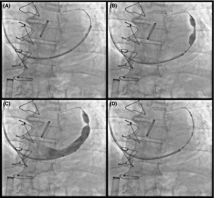
Implantation of a second reducer a few millimeters more distal to the landing zone of the first reducer (A and B), with expansion and the final result (C and D)
References
-
- Knuuti J, Wijns W, Saraste A, et al. 2019 ESC Guidelines for the diagnosis and management of chronic coronary syndromes. Eur Heart J. 2020;41(3):407‐477. - PubMed
-
- Konigstein M, Giannini F, Banai S. The Reducer device in patients with angina pectoris: mechanisms, indications, and perspectives. Eur Heart J. 2018;39(11):925‐933. - PubMed
-
- Tzanis G, Palmisano A, Gallone G, et al. The impact of the coronary sinus reducer upon left ventricular function in patients with refractory angina pectoris. Catheter Cardiovasc Interv. 2020;95:1104‐1108. - PubMed
-
- Palmisano A, Giannini F, Rancoita P, et al. Feature tracking and mapping analysis of myocardial response to improved perfusion reserve in patients with refractory angina treated by coronary sinus Reducer implantation: a CMR study. Int J Cardiovasc Imaging. 2020. ahead of print. 10.1007/s10554-020-01964-9 - DOI - PubMed
Publication types
LinkOut - more resources
Full Text Sources
