Case Reports
. 2021 Jan 11;10(1):2058460120985519.
doi: 10.1177/2058460120985519. eCollection 2021 Jan.

Spontaneous dissolution of a cyst located within the septum pellucidum in a patient with sarcoidosis: a case report

Affiliations
Case Reports

Spontaneous dissolution of a cyst located within the septum pellucidum in a patient with sarcoidosis: a case report

Martina Kastrup Loft et al. Acta Radiol Open. .

Abstract

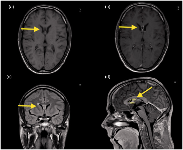
Sarcoidosis is a granulomatous multisystem disease of unknown etiology. Typically, the disease affects the lungs, causing enlargement of the mediastinal lymph nodes, but other organs can be affected. Neurosarcoidosis is reported in 5-10% of the patients. This case represents a 39-year-old male patient diagnosed with lung sarcoidosis. Due to neurological symptoms, a contrast-enhanced cerebral magnetic resonance imaging was performed. Neurosarcoidosis was presented with meningeal enhancement adjacent to a cyst located within the cavum septum pellucidum. The cyst dissolved spontaneously within six months. The finding of a cyst located within the septum pellucidum is rare.

Keywords: MRI; cyst; septum pellucidum; spontaneous dissolution.


Figures

Figure 1.
Figure 1.
MRI images from the first scan performed. Arrows shows the location of the cyst. (a) T1 (axial plane). The cyst contains fluid. (b) T1 including gadolinium (axial plane). Meningeal enhancement adjacent to the cyst. (c) FLAIR sequence (coronal plane). Fluid inside the cyst differs from the CSF. (d) T1 including gadolinium (sagittal plane). Meningeal enhancement adjacent to the cyst.
Figure 2.
Figure 2.
MRI images from the last MRI scan performed. (a) T1 (axial plane). No persisting cyst. (b) T2 (axial plane). No meningeal enhancement. Normal appearance of cerebrum. (c) MRI apparent diffusion coefficient map image (axial plane). Normal appearance of cerebrum. (d) T1 including gadolinium (sagittal plane). No persistent meningeal enhancement.
Figure 3.
Figure 3.
Sagittal cerebrum MRI (T1 including gadolinium) image. (a) Baseline scan. The cyst is present. (b) Two-month of follow-up. The cyst is present. (c) Five-month of follow-up. No visible cyst. (d) 17-month of follow-up. No visible cyst.

References

    1. Voortman M, Drent M, Baughman R. Management of neurosarcoidosis: a clinical challenge. Curr Opin Neurol 2019; 32:475–483. - PMC - PubMed
    1. Fritz D, van de Beek D, Brouwer MC. Clinical features, treatment and outcome in neurosarcoidosis: systematic review and metaanalysis. BMC Neurol 2016; 16:220. - PMC - PubMed
    1. Oteruelo FT. On the cavum septi pellucidi and the cavum vergae. Anat Anz 1986; 162:271–278. - PubMed
    1. Hebel R, Dubaniewicz-Wybieralska M, Dubaniewicz A. Overview of neurosarcoidosis: recent advances. J Neurol 2015; 262:258–267. - PMC - PubMed
    1. Joseph FG, Scolding NJ. Neurosarcoidosis: a study of 30 new cases. J Neurol Neurosurg Psychiatry 2008; 80:297–304. - PubMed

Publication types

LinkOut - more resources