Case Reports
. 2020 Dec 28:2020:8875547.
doi: 10.1155/2020/8875547. eCollection 2020.

Plasmablastic Lymphoma Associated with Adjacent Mature Plasma Cell Population Exhibiting Opposite Light Chain Restriction

Affiliations
Case Reports

Plasmablastic Lymphoma Associated with Adjacent Mature Plasma Cell Population Exhibiting Opposite Light Chain Restriction

Karina Furlan et al. Case Rep Pathol. .

Abstract

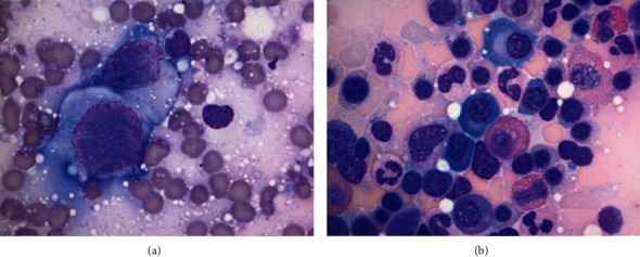
Plasmablastic lymphoma (PBL) is an aggressive high-grade B cell lymphoma, considered a variant of diffuse large B cell lymphoma with approximately 75% mortality within 6-7 months. We describe an unusual case of PBL arising as a maxillary mass in an HIV-negative, nontransplanted 78-year-old female. Histologic examination revealed a diffuse infiltrate of anaplastic appearing cells exhibiting plasmablastic morphology with an adjacent contiguous infiltrate of mature appearing plasma cells. The PBL and mature plasma cell components both demonstrated an immunophenotype of CD20(-), CD38(+), and CD138(+). The two populations differed by the PBL featuring a high proliferation rate by Ki-67 (~95%) with coexpression of both c-MYC and EBV, while the mature plasma cell component featured a low proliferation rate by Ki-67 (~5%) without coexpression of c-MYC or EBV. Kappa/lambda staining demonstrated lambda light chain restriction involving the PBL, while the mature plasma cell infiltrate revealed kappa light chain restriction. Our findings describe the rare association of PBL with a synchronous distinct population of mature plasma cells exhibiting opposite light chain restriction.


Conflict of interest statement

The authors declare that they have no conflicts of interest.

Figures

Figure 1
Figure 1
(a) CT right maxillary sinus mass. (b) Tumor cells forming sheets with a relatively cohesive growth pattern many exhibiting plasmablastic morphology (H&E, 400x). (c) Bizarre/anaplastic forms (H&E, 400x). (d) Abundant mitotic and apoptotic activity (H&E, 200x).
Figure 2
Figure 2
The large tumor cells are positive for MUM1 ((a) immunoperoxidase stain, 40x), partial positive for CD138 ((b) immunoperoxidase stain, 200x), and negative for CD20 ((c) immunoperoxidase stain, 40x) with Epstein–Barr virus-encoded RNA (EBER) transcripts identified by in situ hybridization ((d), 40x).
Figure 3
Figure 3
(a) Immunohistochemical stain for lambda, positive in the PBL (blue arrow) and predominately negative in the mature plasma cell component (red arrow) (100x). (b) Immunohistochemical stain for kappa, positive in the mature plasma cell component (red arrow) and essentially negative in the PBL cells (blue arrow) (100x). (c) Dual immunohistochemical stain for kappa and lambda showing lambda light chain restriction in the PBL cells (blue arrow) and kappa restriction in the mature plasma cell component (red arrow) (100x).
Figure 4
Figure 4
(a) The mature plasma cell population (H&E, 200x). (b) Interface of PBL (blue arrow) and mature plasma cell population (red arrow) (H&E, 100x). (c)–(e) Interface of PBL and mature plasma cell population showing opposite expression of EBER-ISH ((c), 40x), Ki-67 ((d), 40x), and c-MYC ((e), 40x).
Figure 5
Figure 5
Immunoglobulin heavy chain clonality testing by next-generation sequencing. (a) Results from the plasmablastic lymphoma demonstrating two distinct peaks consistent with a biallelic rearranged clone. (b) Results from the mature plasma cell neoplasm with inclusive results due to a low number of reads, but demonstrating an oligoclonal pattern with low-level detection of the biallelic clone found in the PBL population.
Figure 6
Figure 6
(a) Touch preparations from the PBL maxillary mass showing malignant cytology featuring enlarged nuclei, with high nuclear/cytoplasmic ratios, fine open reticular chromatin, and multiple prominent nucleoli (Diff-Quik stain, 1000x). (b) Aspirate smears from the bone marrow biopsy showing mature plasma cells (Wright-Giemsa, 1000x).

References

    1. Delecluse H. J., Anagnostopoulos I., Dallenbach F., et al. Plasmablastic lymphomas of the oral cavity: a new entity associated with the human immunodeficiency virus infection. Blood. 1997;89(4):1413–1420. doi: 10.1182/blood.V89.4.1413. - DOI - PubMed
    1. Montes-Moreno S., Gonzalez-Medina A. R., Rodriguez-Pinilla S. M., et al. Aggressive large B-cell lymphoma with plasma cell differentiation: immunohistochemical characterization of plasmablastic lymphoma and diffuse large B-cell lymphoma with partial plasmablastic phenotype. Haematologica. 2010;95(8):1342–1349. doi: 10.3324/haematol.2009.016113. - DOI - PMC - PubMed
    1. Valera A., Colomo L., Martínez A., et al. ALK-positive large B-cell lymphomas express a terminal B-cell differentiation program and activated STAT3 but lack _MYC_ rearrangements. Modern Pathology. 2013;26(10):1329–1337. doi: 10.1038/modpathol.2013.73. - DOI - PMC - PubMed
    1. Castillo J. J., Bibas M., Miranda R. N. The biology and treatment of plasmablastic lymphoma. Blood. 2015;125(15):2323–2330. doi: 10.1182/blood-2014-10-567479. - DOI - PubMed
    1. Tchernonog E., Faurie P., Coppo P., et al. Clinical characteristics and prognostic factors of plasmablastic lymphoma patients: analysis of 135 patients from the LYSA group. Annals of Oncology. 2017;28(4):843–848. doi: 10.1093/annonc/mdw684. - DOI - PubMed

Publication types