Review
. 2021 Jan 4:12:1-23.
doi: 10.3762/bjnano.12.1. eCollection 2021.

Bio-imaging with the helium-ion microscope: A review

Affiliations
Review

Bio-imaging with the helium-ion microscope: A review

Matthias Schmidt et al. Beilstein J Nanotechnol. .

Abstract

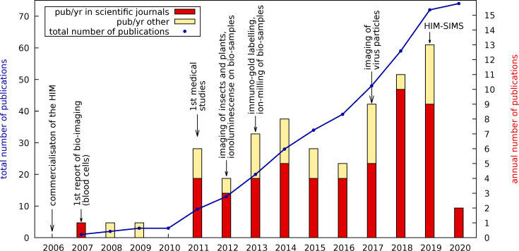
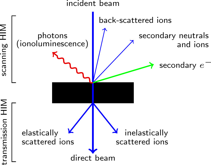
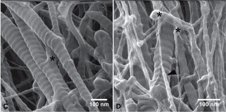
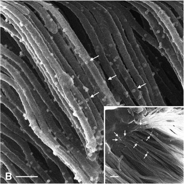
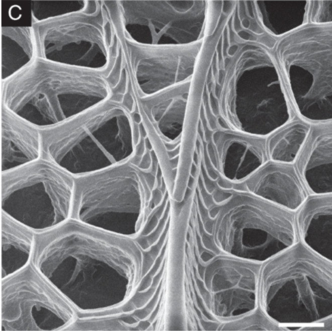
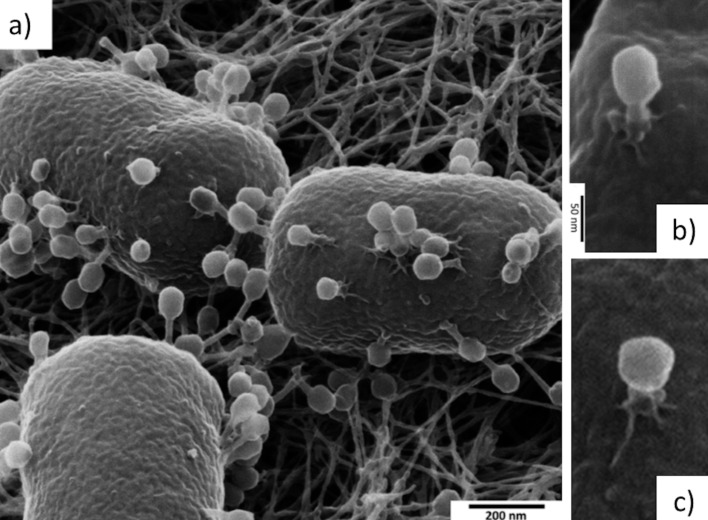
Scanning helium-ion microscopy (HIM) is an imaging technique with sub-nanometre resolution and is a powerful tool to resolve some of the tiniest structures in biology. In many aspects, the HIM resembles a field-emission scanning electron microscope (FE-SEM), but the use of helium ions rather than electrons provides several advantages, including higher surface sensitivity, larger depth of field, and a straightforward charge-compensating electron flood gun, which enables imaging of non-conductive samples, rendering HIM a promising high-resolution imaging technique for biological samples. Starting with studies focused on medical research, the last decade has seen some particularly spectacular high-resolution images in studies focused on plants, microbiology, virology, and geomicrobiology. However, HIM is not just an imaging technique. The ability to use the instrument for milling biological objects as small as viruses offers unique opportunities which are not possible with more conventional focused ion beams, such as gallium. Several pioneering technical developments, such as methods to couple secondary ion mass spectrometry (SIMS) or ionoluminescence with the HIM, also offer the possibility for new and exciting research on biological materials. In this review, we present a comprehensive overview of almost all currently published literature which has demonstrated the application of HIM for imaging of biological specimens. We also discuss some technical features of this unique type of instrument and highlight some of the new advances which will likely become more widely used in the years to come.

Keywords: HIM; HIM-SIMS; bio-imaging; flood gun; helium-ion microscopy; high resolution; ionofluorescense.


Figures

Figure 1
Figure 1
Annual (bars) and accumulated (blue line) numbers of publications on bio-imaging using the helium-ion microscope since its commercialisation. Some important milestones are also indicated. Red bars refer to annual publications in peer-reviewed scientific journals, yellow bars list other publications, such as white papers, Ph.D. theses, and extended conference abstracts.
Figure 2
Figure 2
When the ion beam of the HIM interacts with the sample, primary ions and secondary particles escape the sample and can be detected. The figure schematically depicts particles which are, in principle, detectable in the scanning HIM and, in the case of a sufficiently thin sample, in the transmission HIM.
Figure 3
Figure 3
Charge compensation in the HIM using the flood gun. An uncoated maize root was imaged with different dwell times, td = {0.1,0.5,20} μs, while flood time, tf = 10 μs, image size, 2048 × 2048 pixels, and brightness/contrast settings of the Everhart–Thornley detector were kept constant. The histograms clearly show a shift towards lower pixel intensities with increasing dwell time. No flooding at all (not shown here) resulted in a black image. The field of view is 20 μm. Unpublished data, sample provided by Yalda Davoudpour.
Figure 4
Figure 4
HIM of two Pseudomonas putida biofilms grown on polyvinylchloride coverslips in parallel under exactly the same conditions. Both films were chemically fixed with 2% glutaraldehyde for 2 h at room temperature. Subsequently (A) was dehydrated in a graded ethanol series and dried with hexamethyldisilazane. In contrast, (B) was not dried but the water was substituted with the ionic liquid 1-butyl-3-methylimidazolium tetrafluoroborate according to the protocol published by Golding et al. [69]. Unpublished data, sample provided by Nedal Said.
Figure 5
Figure 5
Rabbit cartilage collagen imaged by HIM. High resolution and depth of field reveal nanoscopic network features such as nanofibrils and their connections (asterisks). Dwell time and resolution are, respectively, 2 μs, 0.90 nm (left) and 0.5 μs, 0.81 nm (right). Reproduced from [70]. Copyright ©2012 The Authors Journal of Microscopy; ©2012 Royal Microscopical Society. Used with permission from Vanden Berg-Foels et al., Helium ion microscopy for high-resolution visualization of the articular cartilage collagen network, Journal of Microscopy, John Wiley and Sons.
Figure 6
Figure 6
Helium-ion micrograph showing an immunogold-labelled (arrows) proximal tubule in a mouse kidney. Scale bar is 200 nm. Reproduced from [15]. Copyright ©2013 Rice et al., distributed under the terms and conditions of the Creative Commons Attribution License CC BY 4.0., https://creativecommons.org/licenses/by/4.0/.
Figure 7
Figure 7
HIM image of a Papilio ulysses butterfly black ground scale. Scale bar is 400 nm. Adapted from [12]. Copyright 2011 ©Wiley Periodicals, Inc. Used with permission from Boden et al., Helium ion microscopy of Lepidoptera scales, Scanning, John Wiley and Sons.
Figure 8
Figure 8
Helium-ion micrographs of the T4 bacteriophage infecting Escherichia coli. (a) Three bacteria with ongoing infection. (b) A higher-resolution image of a single T4 bacteriophage attached on the cell surface. The tail is contracted and the tail fibers are spread out, indicating a genome injection in progress. The icosahedral shape of the head is also apparent. (c) Another individual phage with even more contracted tail. Adapted from [17]. Copyright ©2017 WILEY-VCH Verlag GmbH & Co. KGaA, Weinheim. Used with permission from Leppänen et al., Imaging bacterial colonies and phage-bacterium interaction at sub-nanometre resolution using helium-ion microscopy, Advanced Biosystems, John Wiley and Sons.
Figure 9
Figure 9
Helium-ion micrograph of the predatory bacterium Bdellovibrio bacteriovorus infecting Escherichia coli. The sample was prepared by M. Krüger and N. Said as preliminary work for the study published in [19].
Figure 10
Figure 10
HIM of Toxoplasma gongii inside a vacuole of an infected Rhesus monkey kidney epithelial cell. The close-ups (A) and (B) show the intravacuolar network of tubules formed by the parasite soon after invasion. White arrows point at bifurcating tubules, black arrows point at crossing tubules that do not fuse. Adapted from [83], Journal of Structural Biology, Vol. 191(1), by de Souza et al., “New views of the Toxoplasma gondii parasitophorous vacuole as revealed by Helium Ion Microscopy (HIM)”, pages 76–85, Copyright (2015), with permission from Elsevier.
Figure 11
Figure 11
Microbial mat collected at the Himalayan hot springs at Manikaran imaged with the HIM. Unpublished data from the study of A. Sharma et al. [18].
Figure 12
Figure 12
A biofilm of Chlorella microalgae imaged using HIM. Charge compensation allowed for imaging the biofilm without any metallisation. The exopolymeric polymeric substances between the algal cells are visualised with high contrast owing to the high surface sensitivity of the HIM. The sample was prepared by J. H. Moreno-Osorio within the study published in [46].
Figure 13
Figure 13
Helium-ion micrograph of a twisted stalk produced by a microaerophilic Fe(II)-oxidizing bacterium (unpublished). The white arrow indicates a single bacterium.
Figure 14
Figure 14
Helium-ion micrographs of the predatory nematode Pristionchus pacificus before (a) and after (b) the removal of the membraneous sheath covering the internal tooth structure by Ne-ion milling. Scale bar 5 μm. Adapted from [6], Joens et al., “Helium Ion Microscopy (HIM) for the imaging of biological samples at sub-nanometer resolution.” Licensed under a Creative Commons Attribution-NonCommercial-NoDerivs 3.0 Unported License, http://creativecommons.org/licenses/by-nc-nd/3.0/. Copyright © 2013, with permission from Springer Nature. This figure must not be reproduced or adapted without permission from Springer Nature.
Figure 15
Figure 15
Helium-ion micrographs of sectioned microbiological samples. (a) He-ion-milled cross section of a E. coli with a half-cut bacteriophage on top of it. (b) Ne-ion-milled cross section of a Bdellovibrio-E. coli bdelloblast with visible internal structure and Bdellovibrio progeny penetrating the membrane. Figure 15a adapted from [17], Copyright © 2017 WILEY-VCH Verlag GmbH & Co. KGaA, Weinheim. Used with permission from Leppänen et al., Imaging bacterial colonies and phage-bacterium interaction at sub-nanometer resolution using helium-ion microscopy, Advanced Biosystems, John Wiley and Sons; Figure 15b adapted from [19], Copyright © 2018 WILEY-VCH Verlag GmbH & Co. KGaA, Weinheim. Used with permission from Said et al., Have an Ion on It: The Life-Cycle of Bdellovibrio bacteriovorus Viewed by Helium-Ion Microscopy, Advanced Biosystems, John Wiley and Sons.
Figure 16
Figure 16
Helium-ion micrograph of a Ne-ion-milled section of a E. coli–nanopillared dragonfly wing interface. A partly deformed and stretched membrane is marked as “S”, and the tip of a nanopillar as “T”. Scale bar 200 nm. Adapted with permission from [104]. Copyright 2020 American Chemical Society.

References

    1. Ward B W, Notte J A, Economou N P. J Vac Sci Technol, B: Microelectron Nanometer Struct–Process, Meas, Phenom. 2006;24:2871–2874. doi: 10.1116/1.2357967. - DOI
    1. Ward B, Notte J A, Economou N P. Photonics Spectra. 2007;41:68–70.
    1. Notte J, Ward B, Economou N, Hill R, Percival R, Farkas L, McVey S. AIP Conf Proc. 2007;931:489–496. doi: 10.1063/1.2799423. - DOI
    1. Scipioni L, Stern L A, Notte J, Sijbrandij S, Griffin B. Adv Mater Processes. 2008;166(6):27–30. Product code: ZAMP16606P27.
    1. Carl Zeiss Microscopy, White paper: Zeiss Orion Plus (2008), (not available online anymore).

LinkOut - more resources