

Spontaneous utero-cutaneous fistula between a benign uterine leiomyoma and abdominal skin: A case report
- PMID: 33489783
- PMCID: PMC7809161
- DOI: 10.1016/j.crwh.2020.e00282
Spontaneous utero-cutaneous fistula between a benign uterine leiomyoma and abdominal skin: A case report
Abstract
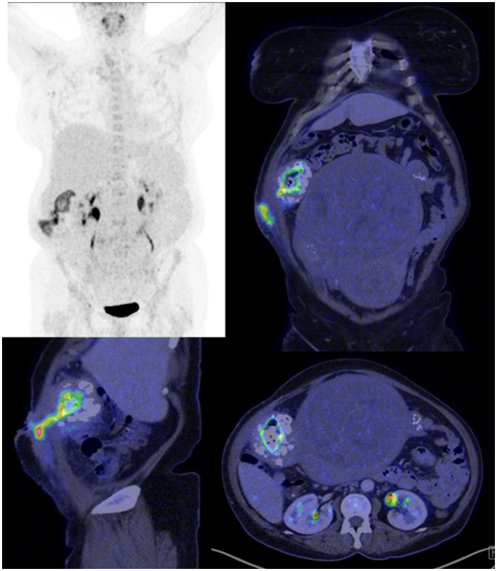
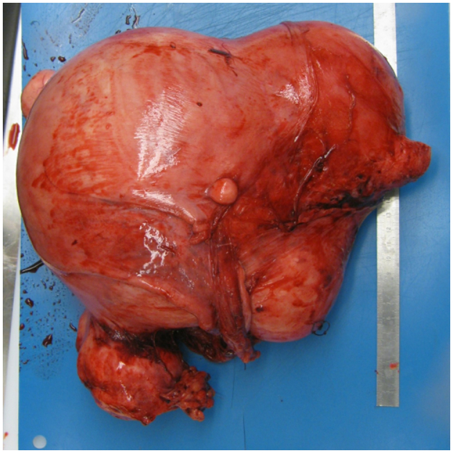
The case of a 56-year-old woman with a fibroid uterus who developed utero-cutaneous fistula is presented. The woman was para 0, had an unremarkable medical history, and had no prior diagnosis of a gynecologic pathology, no operative interventions involving the uterine wall or any other risk factor for fistula. Abdominal examination revealed an abdominal mass with overlying deep, purulent ulceration. 18F-FDG PET/CT scan was consistent with uterine leiomyoma, but a differential diagnosis of sarcoma was considered due to the presence of the fistula, patchy increased FDG uptake of the tumor and several mildly enlarged lymph nodes bilaterally in the inguinal and iliac region. Hysterectomy with bilateral salpingo-oophorectomy was performed. Histological diagnosis was of leiomyoma with focal bizarre atypia, degenerative and metaplastic changes and utero-cutaneous fistula. To the best of our knowledge, this is the first case report describing a benign leiomyoma forming a fistula between the uterus and abdominal surface.
Keywords: Case report; FDG, fluorodeoxyglucose; Leiomyoma; UAE, uterine artery embolization; Utero-cutaneous fistula.
© 2020 The Authors.
Figures

Similar articles
-
Cutaneous malignant melanoma metastatic to a uterine leiomyoma: a case report.J Reprod Med. 2008 Sep;53(9):697-9. J Reprod Med. 2008. PMID: 18839825
-
Incidental Finding of Metastatic Cutaneous Malignant Melanoma at Uterine Leiomyoma, A Thai University Hospital Experience: A Case Report.J Med Assoc Thai. 2015 Apr;98 Suppl 3:S126-31. J Med Assoc Thai. 2015. PMID: 26387400
-
An unusual presentation of a ruptured degenerative fibroid in a perimenopausal woman.BMJ Case Rep. 2014 Dec 9;2014:bcr2014207473. doi: 10.1136/bcr-2014-207473. BMJ Case Rep. 2014. PMID: 25498115 Free PMC article.
-
A case of cotyledonoid leiomyoma and review of the literature.Int J Gynecol Cancer. 2005 Nov-Dec;15(6):1218-21. doi: 10.1111/j.1525-1438.2005.00181.x. Int J Gynecol Cancer. 2005. PMID: 16343218 Review.
-
Uteroenteric fistula resulting from fibroid expulsion after uterine fibroid embolization: case report and review of the literature.Cardiovasc Intervent Radiol. 2012 Oct;35(5):1231-6. doi: 10.1007/s00270-011-0318-4. Epub 2011 Dec 10. Cardiovasc Intervent Radiol. 2012. PMID: 22159908 Review.
Cited by
-
Utero-Cutaneous Fistula in Ruminants: Characterization of the First Cases in Ewes and Cows.Animals (Basel). 2024 Jan 22;14(2):344. doi: 10.3390/ani14020344. Animals (Basel). 2024. PMID: 38275803 Free PMC article.
References
-
- Prato B. Correlation of recurrence rates and times with posttreatment human papillomavirus status in patients treated with loop electrosurgical excision procedure conization for cervical squamous intraepithelial lesions. Int. J. Gynecol. Cancer. 2008;18(1):90–94. doi: 10.1111/j.1525-1438.2007.00965.x. - DOI - PubMed
Publication types
LinkOut - more resources
Full Text Sources
Other Literature Sources
