. 2021 Jan 8:8:591084.
doi: 10.3389/fbioe.2020.591084. eCollection 2020.

Hydroxyapatite Particle Density Regulates Osteoblastic Differentiation Through β-Catenin Translocation

Affiliations

Hydroxyapatite Particle Density Regulates Osteoblastic Differentiation Through β-Catenin Translocation

Otto J Juhl 4th et al. Front Bioeng Biotechnol. .

Abstract

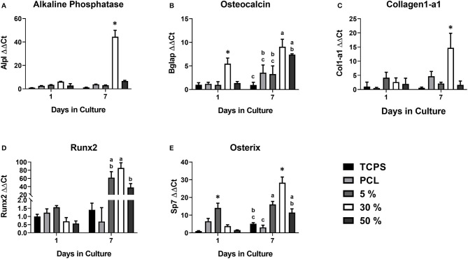
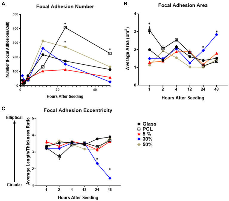
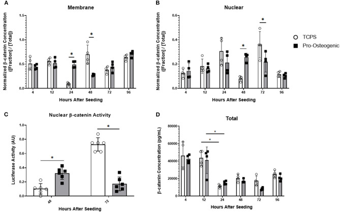
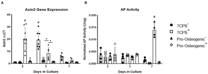
Substrate surface characteristics such as roughness, wettability and particle density are well-known contributors of a substrate's overall osteogenic potential. These characteristics are known to regulate cell mechanics as well as induce changes in cell stiffness, cell adhesions, and cytoskeletal structure. Pro-osteogenic particles, such as hydroxyapatite, are often incorporated into a substrate to enhance the substrates osteogenic potential. However, it is unknown which substrate characteristic is the key regulator of osteogenesis. This is partly due to the lack of understanding of how these substrate surface characteristics are transduced by cells. In this study substrates composed of polycaprolactone (PCL) and carbonated hydroxyapatite particles (HAp) were synthesized. HAp concentration was varied, and a range of surface characteristics created. The effect of each substrate characteristic on osteoblastic differentiation was then examined. We found that, of the characteristics examined, only HAp density, and indeed a specific density (85 particles/cm2), significantly increased osteoblastic differentiation. Further, an increase in focal adhesion maturation and turnover was observed in cells cultured on this substrate. Moreover, β-catenin translocation from the membrane bound cell fraction to the nucleus was more rapid in cells on the 85 particle/cm2 substrate compared to cells on tissue culture polystyrene. Together, these data suggest that particle density is one pivotal factor in determining a substrates overall osteogenic potential. Additionally, the observed increase in osteoblastic differentiation is a at least partly the result of β-catenin translocation and transcriptional activity suggesting a β-catenin mediated mechanism by which substrate surface characteristics are transduced.

Keywords: beta catenin; focal adhesion; mechanotransduction; surface topography; translocation.


Conflict of interest statement

The authors declare that the research was conducted in the absence of any commercial or financial relationships that could be construed as a potential conflict of interest.

Figures

Figure 1
Figure 1
Osteoblastic gene expression on the various PCL/HA substrates. Evaluation of osteoblastic gene expression of (A) alkaline phosphatase (Alpl), (B) osteocalcin (Bglap), (C) collagen1-a1 (Col1-a1), (D) Runx2, and (E) Osterix (Sp7) at 1 and 7 days on the various substrates evaluated in this study. Substrates sharing the same letter denotes a lack of significance at the same time point, *significantly different compared to all other groups at the same time point. n = 3–4 samples with each sample being the average of three replicates.
Figure 2
Figure 2
Focal adhesion number, size, and morphology over time on each of the evaluated substrates. Temporal quantification of focal adhesion (A) number, (B) area, and (C) eccentricity over time on either glass, PCL, 5, 30, or 50% substrates. *p < 0.05 compared to all other groups at the same time point. n = 3 samples with each sample consisting of ~5–75 cells or ~500–5,000 focal adhesions per sample.
Figure 3
Figure 3
β-catenin localization and total β-catenin concentration over 96 h and transcriptional activity at 48 and 72 h in hFOB 1.19 cells cultured on either TCPS or pro-osteogenic substrate. Quantification of normalized β-catenin concentration in either the (A) membrane bound cell fraction, (B) nuclear cell fraction, or (D) total β-catenin concentration. (C) Nuclear β-catenin activity quantified by TOPFLASH activity on either TCPS or pro-osteogenic substrate after 48 h and 72 h. n = 4–6 samples with each sample consisting of two replicates. *p < 0.05.
Figure 4
Figure 4
Inhibition of β-catenin binding to TCF/LEF in hFOB 1.19 cells on TCPS or Pro-Osteogenic Substrates. Evaluation of effect of β-catenin inhibitor, PNU-74564, on (A) Axin2 gene expression and (B) AP Activity in cells cultured in media with inhibitor (+) or without inhibitor (–) on either TCPS or Pro-Osteogenic substrates. *p < 0.05, #p < 0.05 compared to all other groups at same time point. n = 5–6 samples with each sample being the average of 3 replicates or 2 replicates for gene expression and AP activity, respectively.

Similar articles

Cited by

References

    1. Abagnale G., Steger M., Hoa Nguyen V., Hersch N., Sechi A., Joussen S., et al. (2015). Surface topography enhances differentiation of mesenchymal stem cells towards osteogenic and adipogenic lineages. Biomaterials 61, 316–326. 10.1016/j.biomaterials.2015.05.030 - DOI - PubMed
    1. Ahn E. H., Kim Y., Kshitiz An S. S., Afzal J., Lee S., et al. . (2014). Spatial control of adult stem cell fate using nanotopographic cues. Biomaterials 35, 2401–2410. 10.1016/j.biomaterials.2013.11.037 - DOI - PMC - PubMed
    1. Alenghat F. J., Ingber D. E. (2002). Mechanotransduction: all signals point to cytoskeleton, matrix, and integrins. Sci. STKE 2002:pe6. 10.1126/stke.2002.119.pe6 - DOI - PubMed
    1. Baron R., Kneissel M. (2013). WNT signaling in bone homeostasis and disease: from human mutations to treatments. Nat. Med. 19, 179–192. 10.1038/nm.3074 - DOI - PubMed
    1. Bertocchi C., Ravasio A., Ong H. T., Toyama Y., Kanchanawong P. (2019). Mechanical roles of vinculin/β-catenin interaction in adherens junction. bioRxiv 770–735. 10.1101/770735 - DOI

LinkOut - more resources