Review
. 2021 Jan 6:8:609683.
doi: 10.3389/fcell.2020.609683. eCollection 2020.

MiT/TFE Family of Transcription Factors: An Evolutionary Perspective

Affiliations
Review

MiT/TFE Family of Transcription Factors: An Evolutionary Perspective

Martina La Spina et al. Front Cell Dev Biol. .

Abstract

Response and adaptation to stress are critical for the survival of all living organisms. The regulation of the transcriptional machinery is an important aspect of these complex processes. The members of the microphthalmia (MiT/TFE) family of transcription factors, apart from their involvement in melanocyte biology, are emerging as key players in a wide range of cellular functions in response to a plethora of internal and external stresses. The MiT/TFE proteins are structurally related and conserved through evolution. Their tissue expression and activities are highly regulated by alternative splicing, promoter usage, and posttranslational modifications. Here, we summarize the functions of MiT/TFE proteins as master transcriptional regulators across evolution and discuss the contribution of animal models to our understanding of the various roles of these transcription factors. We also highlight the importance of deciphering transcriptional regulatory mechanisms in the quest for potential therapeutic targets for human diseases, such as lysosomal storage disorders, neurodegeneration, and cancer.

Keywords: autophagy; evolution; helix-loop-helix transcription factor 30 (HLH-30); lysosomes; mammalian target of rapamycin (mTOR); microphthalmia-associated transcription factor (MITF); transcription factor E3 (TFE3); transcription factor EB (TFEB).


Conflict of interest statement

The authors declare that the research was conducted in the absence of any commercial or financial relationships that could be construed as a potential conflict of interest.

Figures

FIGURE 1
FIGURE 1
Phylogenetic relationships of the different MiT/TFE family members. Evolutionary comparison of different members of the MiT/TFE protein family represented in a phylogenetic rooted tree generated using MEGA X program (ver. 10.1.8) with a Maximum Likelihood and JTT matrix-based model and 1,000 bootstrap replicates (Jones et al., 1992; Kumar et al., 2018). The tree with the highest log likelihood (–17751.05) is shown. The percentage of trees in which the associated taxa clustered together is shown next to each branch. The tree is drawn to scale, with branch lengths measured in the number of substitutions per site. The scale of the branch lengths is indicated below the tree. The four MiT/TFE members are highlighted in different colors. Mammalian proteins are highlighted in darker colors. Ce, Caenorhabditis elegans; Dm, Drosophila melanogaster; Dr, Danio rerio; Gg, Gallus gallus; Hs, Homo sapiens; L, long; Mm, Mus musculus; P1-3, protein 1–3; Pc, Phasianus colchicus; S, short; Sh, Strigops habroptila; Xt, Xenopus tropicalis.
FIGURE 2
FIGURE 2
Sequence conservation of MiT/TFE transcription factors across species. Clustal Omega multiple sequence alignment of MiT/TFE proteins from Homo sapiens (Hs), Danio rerio (Dr), Drosophila melanogaster (Dm), and Caenorhabditis elegans (Ce). Shaded boxes highlight the degree of conservation of key functional domains between all proteins and species. Arrowheads indicate posttranslational modifications described in human TFEB, showing different degrees of conservation between species. formula image 
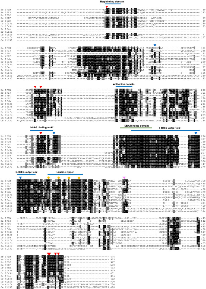
Phosphorylation at serines 3, 138, 142, 211, 462, 463, 467, and 469; formula image Acetylation at lysines 116, 219, 274, and 279; formula image Sumoylation at lysine 346. Diamonds (formula image) point out leucine residues 298, 305, 312, and 319 important for the leucine zipper domain function. Note that the N-terminal sequences of some of the proteins analyzed were omitted due to the figure size constraints.
FIGURE 3
FIGURE 3
MiT/TFE activation is regulated in response to nutrient deprivation. Schematic representation of the mechanism of MiT/TFE transcription factor regulation by nutrient levels in the cell. In nutrient-rich condition, MiT/TFE proteins mammalian target of rapamycin complex 1 (mTORC1) are recruited to the lysosomal membrane through binding to active RagGTPases. Active mTORC1 phosphorylates MiT/TFE proteins at key residues that creates a binding site for the 14-3-3, which sequesters the transcription factors inactive in the cytosol. Under nutrient deprivation conditions, inactive RagGTPases lead to mTORC1 inactivation and MiT/TFE protein dissociation from 14-3-3 as a consequence of their dephosphorylation mediated by the calcium-dependent activation of calcineurin. Nuclear accumulation of MiT/TFE proteins mediates the activation of a transcriptional network that promotes autophagy, lysosomal biogenesis, and increased lysosomal degradation. Upon nutrient replenishment conditions, MiT/TFE proteins nuclear export is regulated by mTOR-dependent phosphorylation and binding to CRM1/Exportin-1. The question marks denote that there is no direct evidence available to support the indicated processes for some of the MiT/TFE family members. CRM1, chromosomal maintenance 1; NPC, nuclear pore complex; P, phosphorylation; Rheb, Ras homolog enriched in brain; v-ATPase, vacuolar-type H+-ATPase.
FIGURE 4
FIGURE 4
Various stress conditions activate MiT/TFE transcription factors. In C. elegans and mammalian cells, different internal and external stress conditions such as DNA damage, protein aggregate accumulation in endo-lysosomal compartments, endoplasmic reticulum (ER) stress, oxidative stress, and mitochondrial dysfunction lead to the activation of MiT/TFE transcription factors. In response to stress, these transcription factors upregulate the expression of a gene network involved in lysosomal function and autophagy flux to promote organismal survival. However, under severe stress conditions, MiT/TFE transcription factors may lead to the expression of apoptotic genes, promoting cell death.
FIGURE 5
FIGURE 5
MiT/TFE transcription factors modulate lysosome, autophagosome, and mitochondrial biogenesis in response to stress. Summary of organelle biogenesis induction in different animal models controlled by the MiT/TFE proteins. In most of the animal models, the activation of the MiT/TFE proteins in response to stress conditions induces an upregulation of genes involved in lysosomal biogenesis, autophagy, and mitochondrial biogenesis. The question marks signify that there are no data available to support organelle biogenesis induction in the indicated animal models; however, it is likely that these processes may take place based on the highly conserved functions between the different MiT/TFE family members across species.
FIGURE 6
FIGURE 6
Key metabolic pathways are regulated by MiT/TFE transcription factors under energy demand conditions. (A) MiT/TFE proteins regulate cellular energy state and metabolism in mammals. Activated and overexpressed (oe) TFEB and TFE3 regulate lipid metabolism and insulin signaling pathways in metabolic organs such as liver, skeletal muscle, and adipose tissue. By upregulating genes involved in autophagy/lipophagy, insulin signaling, and degradation of lipids and in the utilization of glucose to promote glycogen synthesis, these transcription factors play an essential role in reducing diabetes and obesity in mice. (B) In D. melanogaster, under nutrient deprivation conditions, Mitf overexpression (oe) induces a reduction in lipid droplet size through the activation of autophagy and increase in lysosomal activity. (C) In fasted C. elegans, helix-loop-helix transcription factor 30 (HLH-30) upregulates the expression of lysosomal lipases (LIPL-1 and LIPL-3) and autophagy genes controlling lipid mobilization via lipolysis. Also, the activation of HLH-30 is central for the survival response to conditions of fasting-refeeding via TOR regulation. ↺ indicates autoregulatory loop. ↑↓ indicate upregulation and downregulation, respectively. BAT, brown adipose tissue; WAT, white adipose tissue.
FIGURE 7
FIGURE 7
The stress response to pathogen infection and inflammation is modulated by MiT/TFE transcription factors. (A) In C. elegans, the activation of helix-loop-helix transcription factor 30 (HLH-30) is mediated by S. aureus and pore-forming toxin Cry5B. (B) In mammalian macrophages, TFEB and TFE3 activation is regulated by many different stimuli. Activated TFEB and TFEB3 upregulate the expression of genes involved in autophagy/lysosome-related processes and inflammatory cytokine/chemokine production. AKT, v-Akt murine thymoma viral oncogene homolog; AMPK, AMP-activated protein kinase; BECN1, beclin-1; FLCN, folliculin; HIV, human immunodeficiency virus; HMOX1, heme oxygenase 1; IFNγ, interferon γ; IFNγR, interferon γ receptor; LPS, lipopolysaccharide; Mtb, M. tuberculosis; Nef, negative regulatory factor; PKCα, protein kinase C alpha; PKD, protein kinase D; PLC, phospholipase C; TLR4, Toll-like receptor 4; TLR8, Toll-like receptor 8.
FIGURE 8
FIGURE 8
Longevity and survival are promoted by MiT/TFE transcription factors. The MiT/TFE transcription factors control longevity and survival, inducing correct development, promoting healthy aging, and preventing cognitive decline. In C. elegans, helix-loop-helix transcription factor 30 (HLH-30) regulates life span extension in starvation-induced adult reproductive diapause conditions. During development, MiT/TFE proteins regulate differentiation of the eye and epidermis pigmented cells in D. melanogaster and proper vertebra formation during embryogenesis in zebrafish notochord. In addition, MiT/TFE proteins repress myelinization and induce autophagy and lysosome integrity to face aging neuronal decline in neurodegenerative lysosomal storage disorders and aberrant protein accumulation.
FIGURE 9
FIGURE 9
MiT/TFE transcription factors regulate cell fate and lineage decision. (A) In C. elegans, helix-loop-helix transcription factor 30 (HLH-30) regulates entry and recovery of adult reproductive quiescence state induced by starvation. (B) MiT/TFE transcription factors are activated under DNA damage, and depending on the severity of the damage, they can play a dual role inducing apoptosis and cell death or cell survival. (C) The MiT/TFE transcription factors regulate cell cycle and proliferation. TFEB can promote a quiescent state in neural stem cell by blocking pro-proliferative inputs. In addition, these transcription factors can promote proliferation rate in the liver and vascular system; however, they can also induce tumorigenesis.

References

    1. Ahmad G., Amiji M. M. (2017). Cancer stem cell-targeted therapeutics and delivery strategies. Expert. Opin. Drug. Deliv. 14 997–1008. 10.1080/17425247.2017.1263615 - DOI - PubMed
    1. Awad O., Sarkar C., Panicker L. M., Miller D., Zeng X., Sgambato J. A., et al. (2015). Altered TFEB-mediated lysosomal biogenesis in Gaucher disease iPSC-derived neuronal cells. Hum. Mol. Genet. 24 5775–5788. 10.1093/hmg/ddv297 - DOI - PubMed
    1. Bahrami A., Bianconi V., Pirro M., Orafai H. M., Sahebkar A. (2020). The role of TFEB in tumor cell autophagy: diagnostic and therapeutic opportunities. Life Sci. 244:117341. 10.1016/j.lfs.2020.117341 - DOI - PubMed
    1. Ballabio A., Bonifacino J. S. (2020). Lysosomes as dynamic regulators of cell and organismal homeostasis. Nat. Rev. Mol. Cell Biol. 21 101–118. 10.1038/s41580-019-0185-4 - DOI - PubMed
    1. Bennett C. F., Kwon J. J., Chen C., Russell J., Acosta K., Burnaevskiy N., et al. (2017). Transaldolase inhibition impairs mitochondrial respiration and induces a starvation-like longevity response in Caenorhabditis elegans. PLoS Genet. 13:e1006695. 10.1371/journal.pgen.1006695 - DOI - PMC - PubMed

LinkOut - more resources