. 2021 Jan 6:8:609708.
doi: 10.3389/fcell.2020.609708. eCollection 2020.

Structural Roles for the Juxtamembrane Linker Region and Transmembrane Region of Synaptobrevin 2 in Membrane Fusion

Affiliations

Structural Roles for the Juxtamembrane Linker Region and Transmembrane Region of Synaptobrevin 2 in Membrane Fusion

Yaru Hu et al. Front Cell Dev Biol. .

Abstract

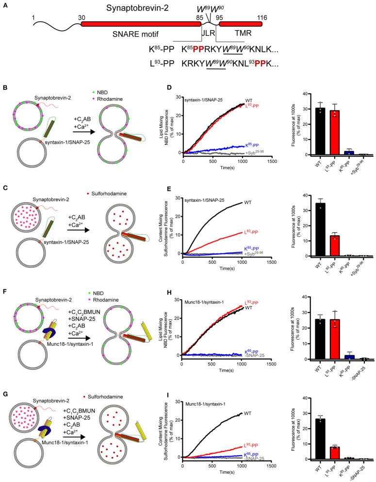
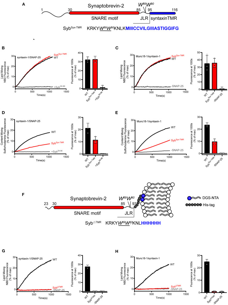
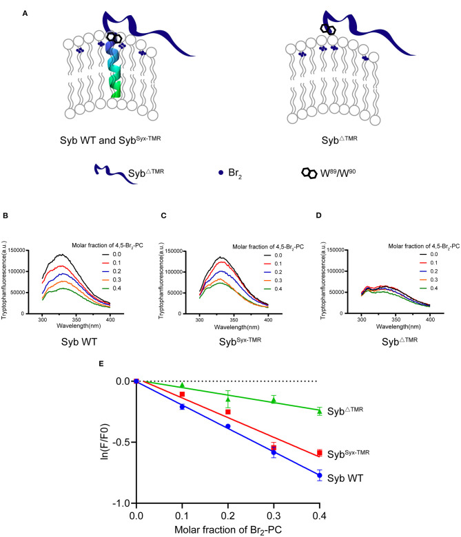
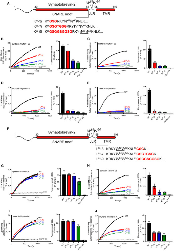
Formation of the trans-SNARE complex is believed to generate a force transfer to the membranes to promote membrane fusion, but the underlying mechanism remains elusive. In this study, we show that helix-breaking and/or length-increasing insertions in the juxtamembrane linker region of synaptobrevin-2 exert diverse effects on liposome fusion, in a manner dependent on the insertion position relative to the two conserved tryptophan residues (W89/W90). Helical extension of synaptobrevin-2 to W89/W90 is a prerequisite for initiating membrane merger. The transmembrane region of synaptobrevin-2 enables proper localization of W89/W90 at the membrane interface to gate force transfer. Besides, our data indicate that the SNARE regulatory components Munc18-1 and Munc13-1 impose liposome fusion strong demand on tight coupling between the SNARE motif and the transmembrane region of synaptobrevin-2.

Keywords: Munc13; Munc18; SNARE complex assembly; membrane fusion; synaptobrevin-2.


Conflict of interest statement

The authors declare that the research was conducted in the absence of any commercial or financial relationships that could be construed as a potential conflict of interest.

Figures

Figure 1
Figure 1
Fusion affected by disrupting helical continuity of the synaptobrevin-2. (A) Domain structure of full length wild-type synaptobrevin-2 (WT) and its mutants with two-proline insertions after K85 (K85-PP) or L93 (L93-PP) in the JLR. (B,C) Scheme of lipid mixing (B) and content mixing assay (C) between syntaxin-1/SNAP-25 and synaptobrevin-2 liposomes in the presence of C2AB fragment and 1 mM Ca2+. Note that the syntaxin-1 SNARE motif (H3, residues 183–288) was used here to form the syntaxin-1/SNAP-25 complex. (D,E) Lipid (D) and content mixing (E) of synaptobrevin-2 WT, K85-PP, and L93-PP liposomes with syntaxin-1/SNAP-25 liposomes. (F,G) Scheme of lipid mixing (F) and content mixing assay (G) between Munc18-1/syntaxin-1 (full length, residues 1–288) and synaptobrevin-2 liposomes in the presence of the Munc13-1 C1-C2B-MUN fragment, SNAP-25, C2AB fragment, and 1 mM Ca2+. (H,I) Lipid (H) and content mixing (I) of synaptobrevin-2 WT, K85-PP, and L93-PP liposomes with Munc18-1/syntaxin-1 liposomes. Representative traces came from one of three independent experiments. Bars on the right panel in (D,E,H,I) are means ± SDs, n = 3.
Figure 2
Figure 2
Fusion affected by substitution or deletion of the synaptobrevin-2 TMR. (A) Domain structure of chimeric synaptobrevin-2 with TMR substituted by syntaxin-1 TMR (residues 267–288) (SybSyx−TMR). (B,C) Lipid mixing between synaptobrevin-2 WT, SybSyx−TMR liposomes and liposomes reconstituted with syntaxin-1/SNAP-25 (B) or Munc18-1/syntaxin-1 (C). (D,E) Content mixing between synaptobrevin-2 WT, SybSyx−TMR liposomes, and liposomes reconstituted with syntaxin-1/SNAP-25 (D) or Munc18-1/syntaxin-1 (E). (F) Domain structure of TMR deletion mutant (SybΔTMR) with C-teminal 6-Histidine tag linked to DGS-NTA-containing liposome. (G,H) Lipid mixing between synaptobrevin-2 WT and SybΔTMR liposomes and liposomes reconstituted with syntaxin-1/SNAP-25 (G) and Munc18-1/syntaxin-1 (H). Representative traces came from one of three independent experiments. Bars on the right panel in (B–E,G,H) are Means ± SDs, n = 3.
Figure 3
Figure 3
W89/W90 of TMR anchored synaptobrevin-2 are resided at the membrane-water interface. (A) Schematic diagrams of lipid quenching assay showing that fluorescence of embedded tryptophans in TMR anchored synaptobrevin-2 were quenched by lipid quencher 4,5-Br2-PC. (B–D) Quenching of tryptophan fluorescence by different molar fraction of 4,5-Br2-PC in synaptobrevin-2 WT (B), SybSyx−TMR (C), and SybΔTMR (D) liposomes. The samples were excited at 285 nm, and the emission spectra were collected in the range of 300–400 nm. (E) Quantification of the fluorescent intensities in (B–D) of synaptobrevin-2 WT (solid blue circles), SybSyx−TMR (solid red squares), and SybΔTMR (solid green triangles) liposome. The total fluorescence intensity (F) was calculated by integrating the intensity in the emission spectral range. (F0) represents the fluorescent intensity in the absence of 4,5-Br2-PC, ln (F/F0) is plotted against the 4,5-Br2-PC molar fraction. Linear regression was performed by Prism 6.01. Bars in (E) are presented as Means ± SDs, n = 3.
Figure 4
Figure 4
Fusion affected by extension of the length or flexibility of the synaptobrevin-2 JLR. (A) Domain structure of full length synaptobrevin-2 with 3, 7, 9 GSG insertions after K85 (K85-3i, K85-7i, K85-9i) in the JLR. (B,C) Lipid mixing (B) and content mixing (C) between synaptobrevin-2 WT, K85-3i, K85-7i, and K85-9i liposomes and syntaxin-1/SNAP-25 liposome. (D,E) Lipid mixing (D) and content mixing (E) between synaptobrevin-2 WT, K85-3i, K85-7i, and K85-9i liposomes and Munc18-1/syntaxin-1 liposome. (F) Domain structure of full-length synaptobrevin-2 with 3, 7, 9 GSG insertions after L93 (L93-3i, L93-7i, L93-9i) in the JLR. (G,H) Lipid mixing (G) and content mixing (H) between synaptobrevin-2 WT, L93-3i, L93-7i, and L93-9i liposomes and syntaxin-1/SNAP-25 liposome. (I,J) Lipid mixing (I) and content mixing (J) between synaptobrevin-2 WT, L93-3i, L93-7i, and L93-9i liposomes and Munc18-1/syntaxin-1 liposome. Representative traces came from one of three independent experiments. Bars on the right panel in (B–E,G–J) are Means ± SDs, n = 3.

References

    1. Bao H., Goldschen-Ohm M., Jeggle P., Chanda B., Edwardson J. M., Chapman E. R. (2016). Exocytotic fusion pores are composed of both lipids and proteins. Nat. Struct. Mol. Biol. 23, 67–73. 10.1038/nsmb.3141 - DOI - PMC - PubMed
    1. Borisovska M., Schwarz Y. N., Dhara M., Yarzagaray A., Hugo S., Narzi D., et al. . (2012). Membrane-proximal tryptophans of synaptobrevin II stabilize priming of secretory vesicles. J. Neurosci. 32, 15983–15997. 10.1523/JNEUROSCI.6282-11.2012 - DOI - PMC - PubMed
    1. Bowen M., Brunger A. T. (2006). Conformation of the synaptobrevin transmembrane domain. Proc. Natl. Acad. Sci. U.S.A. 103, 8378–8383. 10.1073/pnas.0602644103 - DOI - PMC - PubMed
    1. Bretou M., Anne C., Darchen F. (2008). A fast mode of membrane fusion dependent on tight SNARE zippering. J. Neurosci. 28, 8470–8476. 10.1523/JNEUROSCI.0860-08.2008 - DOI - PMC - PubMed
    1. Brewer K. D., Li W., Horne B. E., Rizo J. (2011). Reluctance to membrane binding enables accessibility of the synaptobrevin SNARE motif for SNARE complex formation. Proc. Natl. Acad. Sci. U.S.A. 108, 12723–12728. 10.1073/pnas.1105128108 - DOI - PMC - PubMed

LinkOut - more resources