Review
. 2020 Nov 27;8(1):1-16.
doi: 10.15698/mic2021.01.739.

Milestones in Bacillus subtilis sporulation research

Affiliations
Review

Milestones in Bacillus subtilis sporulation research

Eammon P Riley et al. Microb Cell. .

Abstract

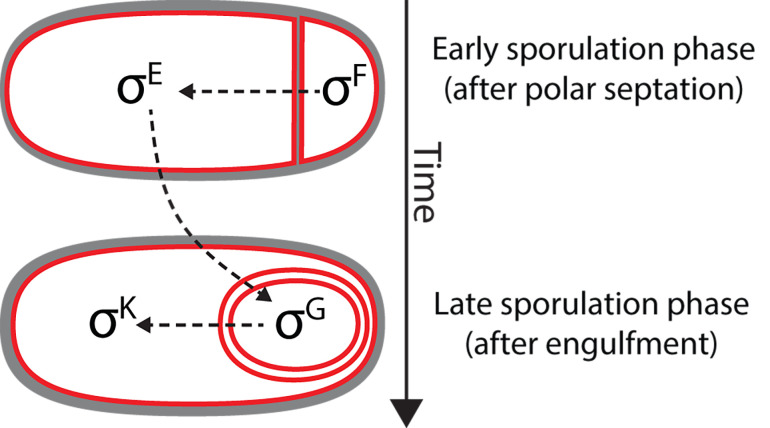
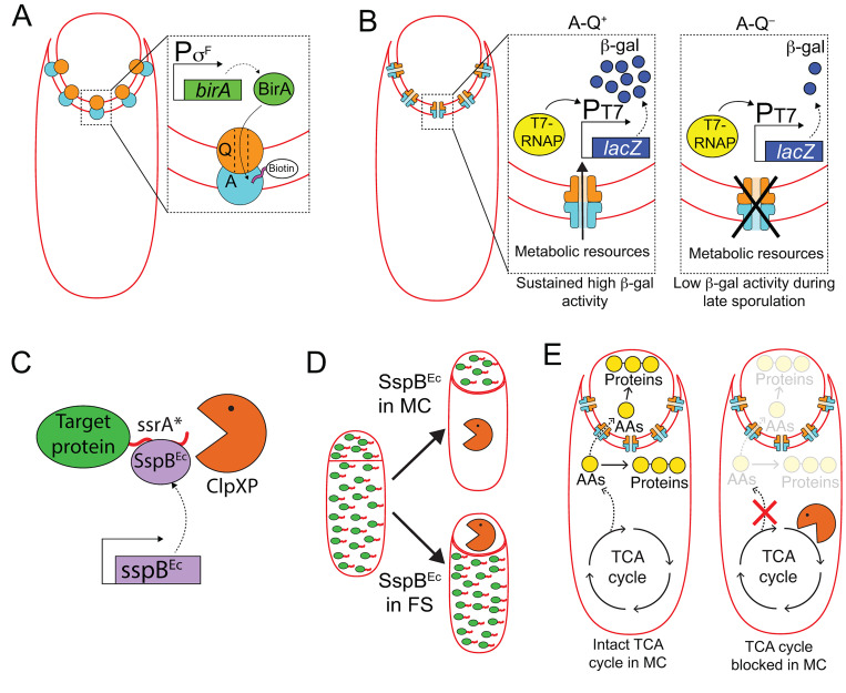
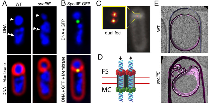
Endospore formation has been a rich field of research for more than a century, and has benefited from the powerful genetic tools available in Bacillus subtilis. In this review, we highlight foundational discoveries that shaped the sporulation field, from its origins to the present day, tracing a chronology that spans more than one hundred eighty years. We detail how cell-specific gene expression has been harnessed to investigate the existence and function of intercellular proteinaceous channels in sporulating cells, and we illustrate the rapid progress in our understanding of the cell biology of sporulation in recent years using the process of chromosome translocation as a storyline. Finally, we sketch general aspects of sporulation that remain largely unexplored, and that we envision will be fruitful areas of future research.

Keywords: Bacillus subtilis; SpoIIIA; SpoIIIE; SpoIIQ; genetics; sporulation; sporulation history.


Conflict of interest statement

Conflict of interest: The authors declare no conflict of interest.

Figures

Figure 1
Figure 1. FIGURE 1: Early depictions of sporulation and germination in Bacillus spp. In one of his most well-known studies [6], Robert Koch investigated the etiology of anthrax.
(A) Koch followed the sporulation process in B. anthracis. He placed a slice of spleen containing the Bacillus into cow sera or aqueous humor, and incubated the specimens at 35-37°C in an incubator that he himself had constructed. During the incubation, he observed the cells growing into string-like structures. After 10-15 h, these strings contained light refracting bodies, which he identified as spores. Koch depicted the structures in this sketch and likened them to fragile strings of pearls. The strings gradually decomposed, the spores were released and sank to the bottom of the droplet where they accumulated and could be kept for weeks. (B) Koch followed spore germination and outgrowth, and determined that spores can form viable cells. He fixed dried spores on a slide and incubated the sample with aqueous humor. After 3-4 hours, he observed various stages of germination and outgrowth, which are depicted in Cohn's sketch. Koch described spores as egg-shaped structures, enclosed by a thin layer of protoplasm that he called bright matter. He hypothesized that during outgrowth this bright matter stretched and became the vegetative cell, whereas the spore remained at one cell pole, lightened, and shrank, before it dissociated and disappeared. He proposed that the spore core consists of an oily substance necessary for the cell to resume vegetative growth.
Figure 2
Figure 2. FIGURE 2: Sporulation stages in B. subtilis.
Figure 3
Figure 3. FIGURE 3: Developmental loci in B. subtilis.
Diagram of the sporulation-germination cycle, with the different classes of developmental loci noted (in parentheses). The spo loci are required for spore formation, but not for vegetative growth. The ger loci are required for proper spore germination. After germination, metabolism is reactivated and the spore transforms into a growing vegetative cell in a process called outgrowth, which is thought to depend on the same pathways that control vegetative growth. The cot and ssp loci were identified after the isolation and characterization of coat and small acid-soluble proteins (SASPs), respectively, from spores. In addition to the these categories, there are hundreds of developmentally-regulated genes whose roles in sporulation and germination are not yet understood. Membranes, red; DNA, blue; cell wall, gray; spore cortex, light gray; spore coat, black; SASPs, green circles.
Figure 4
Figure 4. FIGURE 4: Genetic strategy to identify the cell in which a spo gene is required.
A Spo strain with a mutation in a known spo gene (yellow star) is depicted in Stage I (see Fig. 2) with its two chromosomes, one that will be packed into the forespore (upper chromosome) and one that will remain in the mother cell and be destroyed when the mother cell lyses (lower chromosome). The strain is transformed with genomic DNA from a Spo+ strain at the onset of sporulation. Transformed chromosomes are drawn in purple, non-transformed chromosomes, in blue. Either or both chromosomes are capable of being transformed to spo+. If the spo gene is required in the forespore (left panel), transformation of the forespore chromosome to spo+ rescues the process and generates spores that can germinate and go on to sporulate again (left). But transformation of the mother-cell chromosome to spo+ with no accompanying transformation of the forespore chromosome leaves the forespore chromosome spo so the cells cannot complete the process, and no spores are produced (center). Hence only Spo+ spores are generated if the spo gene is required in the forespore. If the spo gene is required in the mother cell (right panel), transformation of the mother-cell chromosome to spo+ rescues the process (center and right), but only if the forespore chromosome is transformed as well will the spores that are produced be able to germinate into cells that can go on to sporulate again (right). Hence, both Spo+ and Spo spores can be generated if the spo gene is required in the mother cell.
Figure 5
Figure 5. FIGURE 5: Cascade of sporulation-specific σ factors in B. subtilis.
Immediately after polar septation, σF is activated in the forespore. σF activation triggers an intercompartmental signaling cascade that leads to the activation of σE in the mother cell. Together σF and σE control gene expression during engulfment. Roughly coincident with the completion of engulfment, σF is replaced by σG in the forespore, which leads to the subsequent activation of σK in the mother cell. σG and σK control gene expression in the forespore and the mother cell, respectively, after engulfment.
Figure 6
Figure 6. FIGURE 6: Harnessing cell-specific gene expression to study the function of A-Q complexes.
(A) The forespore protein SpoIIQ (Q, orange circle) and the mother-cell proteins encoded in the spoIIIA operon (A, light blue circle), form trans-envelope complexes that bridge the forespore and mother-cell membranes during engulfment. Zoomed in panel: Biotin ligase (BirA, green) produced in the forespore is able to biotinylate a biotin acceptor peptide (pink) fused to the extracellular domain of the A protein, SpoIIIAH [111], indicating that A-Q complexes are channels (see panel B), and that the channel pore is open on the forespore side and large enough for BirA to reach the extracellular domain of SpoIIIAH. (B) The activity of a heterologous RNA polymerase (T7 RNAP, yellow) produced in the forespore under σF control is monitored by the accumulation of β-galactosidase (β-gal, dark blue circles) produced through the expression of a lacZ gene under the control of a T7 RNAP-dependent promoter. In the presence of A-Q channels (A-Q+, left zoomed in panel), sustained β-galactosidase activity is detected throughout sporulation. In the absence of A-Q channels (A-Q, right zoomed in panel), β-galactosidase activity drops at later sporulation stages. Camp and Losick [112] proposed that A-Q channels constitute feeding tubes through which the mother cell transfers metabolic resources to the forespore to maintain biosynthetic activities at late sporulation stages. (C) The ssrA*/SspBEc inducible protein degradation system [114]. SspBEc (purple) binds to ssrA* (red) fused to the C-terminus of target proteins (green), and delivers the target proteins to the endogenous B. subtilis protease ClpXP (orange pacman) for degradation. (D) Cell-specific degradation of target proteins during sporulation is achieved by expressing sspBEc from mother cell- or forespore-specific promoters [113]. Target proteins represented by green ovals tagged with ssrA (red line); degradation represented by orange pacman. (E) Left cell: Mother-cell TCA cycle provides metabolic precursors, such as amino acids (AAs, yellow circles), to support protein synthesis in both the mother cell and the forespore [115]. Mother-cell metabolic precursors could be transported to the forespore via A-Q channels, in keeping with the feeding tube model. Right cell: Degradation of TCA cycle enzymes in the mother cell blocks protein synthesis in both the mother cell and the forespore [115].
Figure 7
Figure 7. FIGURE 7: Progress in our understanding of SpoIIIE-mediated chromosome translocation.
(A) Fluorescence microscopy images of a wild-type (WT, left) and of a spoIIIE mutant (spoIIIE, right) strain of B. subtilis. The upper panels show the DNA stained with DAPI (blue), and the lower panels the overlay of DAPI-stained DNA and the membranes stained with FM 4-64 (red). In the upper panel, forespores are indicated by single arrowheads, and mother cells by double arrowheads. Wild-type forespores contain a complete chromosome. However, forespores of spoIIIE mutant strains contain only ~30% of a chromosome, and the rest remains trapped in the mother cell. (B) Fluorescence microscopy image of a sporulating cell producing a SpoIIIE-GFP fusion during chromosome translocation. The upper panel shows the DNA stained with DAPI (blue) and the GFP signal, and the lower panel shows in addition the membranes stained with FM 4-64 (red). SpoIIIE forms a focus (green) at septal midpoint, where the chromosome is trapped. (C) SpoIIIE visualized by super-resolution microscopy (PALM) in living cells with thicker polar septa. SpoIIIE forms two foci (dual foci), which are separated by a distance equivalent to the septal thickness, indicating that one cluster is present at one side of the septum and the other at the opposite side. Reproduced from [101]. (D) Model for the organization and function of the SpoIIIE translocation complex at the septal midpoint. SpoIIIE forms two side-by-side channels spanning both septal membranes (red lines), thereby allowing the simultaneous transport of both arms of the chromosome from the mother cell (MC) to the forespore (FS). Translocation is powered by SpoIIIE motor domains at the mother-cell side of the septum (green circles), which are activated to export the chromosome to the forespore. Forespore motor domains (red circles) remain inactive. (E) Cryo-electron microscopy of a wild-type sporulating cell (top), and of a spoIIIE mutant (bottom). Membranes are annotated as follows: forespore membrane, pink; mother-cell membrane, purple. Chromosome translocation is required to maintain the shape of the forespore. In the absence of chromosome translocation, the forespore appears deflated. Reproduced with permission from [100].

References

    1. Cano R, Borucki M. Revival and identification of bacterial spores in 25- to 40-million-year-old Dominican amber. Science. 1995;268(5213):1060–1064. doi: 10.1126/science.7538699. - DOI - PubMed
    1. Crichton M. Jurassic Park, 1st ed. Alfred A. Knopf. 1990.
    1. D'Amelio E, Gentile B, Lista F, D'Amelio R. Historical evolution of human anthrax from occupational disease to potentially global threat as bioweapon. Environ Int. 2015;85:133–146. doi: 10.1016/j.envint.2015.09.009. - DOI - PubMed
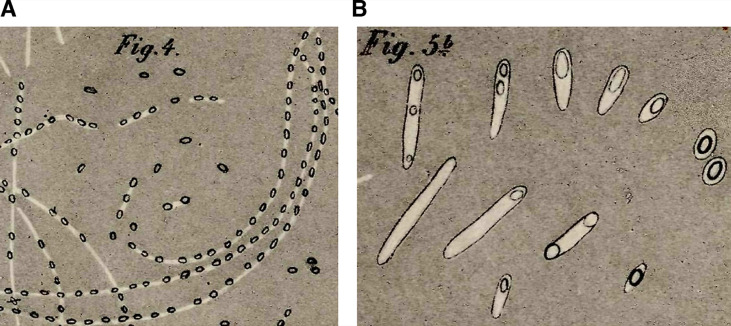
    1. Ehrenberg CG. Die Infusionsthierchen als vollkommene Organismen. Verlag Von Leopold Voss, Leipzig. 1838.
    1. Cohn F. Untersuchungen über Bakterien IV. Beiträge zur Biologie der Bazillen. Beitrage zur Biol der Pflanz. 1876. pp. 249–276.

LinkOut - more resources