

Optical Coherence Tomography (Angiography) Biomarkers in the Assessment and Monitoring of Diabetic Macular Edema
- PMID: 33490283
- PMCID: PMC7790580
- DOI: 10.1155/2020/6655021
Optical Coherence Tomography (Angiography) Biomarkers in the Assessment and Monitoring of Diabetic Macular Edema
Abstract
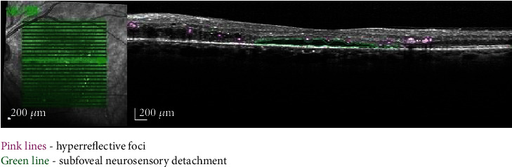
Retinopathy is one of the most severe diabetes-related complications, and macular edema is the major cause of central vision loss in patients with diabetes mellitus. Significant progress has been made in recent years in optical coherence tomography and angiography technology. At the same time, various parameters have been attributed the role of biomarkers creating the frame for new monitoring and treatment strategies and offering new insights into the pathogenesis of diabetic retinopathy and diabetic macular edema. In this review, we gathered the results of studies that investigated various specific OCT (angiography) parameters in diabetic macular edema, such as central subfoveal thickness (CST), cube average thickness (CAT), cube volume (CV), choroidal thickness (CT), retinal nerve fiber layer (RNFL), retinal thickness at the fovea (RTF), subfoveal choroidal thickness (SFCT), central macular thickness (CMT), choroidal vascularity index (CVI), total macular volume (TMV), central choroid thickness (CCT), photoreceptor outer segment (PROS), perfused capillary density (PCD), foveal avascular zone (FAZ), subfoveal neuroretinal detachment (SND), hyperreflective foci (HF), disorganization of the inner retinal layers (DRIL), ellipsoid zone (EZ), inner segment/outer segment (IS/OS) junctions, vascular density (VD), deep capillary plexus (DCP), and superficial capillary plexus (SCP), in order to provide a synthesis of biomarkers that are currently used for the early diagnosis, assessment, monitoring, and outlining of prognosis.
Copyright © 2020 Corina-Iuliana Suciu et al.
Conflict of interest statement
The authors declare that there is no conflict of interest regarding the publication of this paper.
Figures


References
Publication types
MeSH terms
Substances
LinkOut - more resources
Full Text Sources
Medical
Research Materials
Miscellaneous
