

Automated Contouring of Contrast and Noncontrast Computed Tomography Liver Images With Fully Convolutional Networks
- PMID: 33490720
- PMCID: PMC7807136
- DOI: 10.1016/j.adro.2020.04.023
Automated Contouring of Contrast and Noncontrast Computed Tomography Liver Images With Fully Convolutional Networks
Abstract
Purpose: The deformable nature of the liver can make focal treatment challenging and is not adequately addressed with simple rigid registration techniques. More advanced registration techniques can take deformations into account (eg, biomechanical modeling) but require segmentations of the whole liver for each scan, which is a time-intensive process. We hypothesize that fully convolutional networks can be used to rapidly and accurately autosegment the liver, removing the temporal bottleneck for biomechanical modeling.
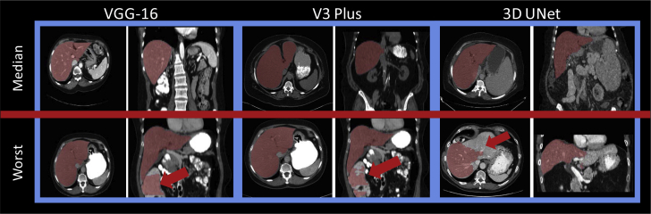
Methods and materials: Manual liver segmentations on computed tomography scans from 183 patients treated at our institution and 30 scans from the Medical Image Computing & Computer Assisted Intervention challenges were collected for this study. Three architectures were investigated for rapid automated segmentation of the liver (VGG-16, DeepLabv3 +, and a 3-dimensional UNet). Fifty-six cases were set aside as a final test set for quantitative model evaluation. Accuracy of the autosegmentations was assessed using Dice similarity coefficient and mean surface distance. Qualitative evaluation was also performed by 3 radiation oncologists on 50 independent cases with previously clinically treated liver contours.
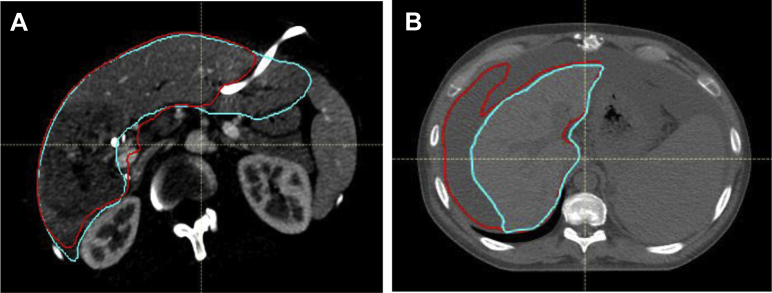
Results: The mean (minimum-maximum) mean surface distance for the test groups with the final model, DeepLabv3 +, were as follows: μContrast(N = 17): 0.99 mm (0.47-2.2), μNon_Contrast(N = 19)l: 1.12 mm (0.41-2.87), and μMiccai(N = 30)t: 1.48 mm (0.82-3.96). The qualitative evaluation showed that 30 of 50 autosegmentations (60%) were preferred to manual contours (majority voting) in a blinded comparison, and 48 of 50 autosegmentations (96%) were deemed clinically acceptable by at least 1 reviewing physician.
Conclusions: The autosegmentations were preferred compared with manually defined contours in the majority of cases. The ability to rapidly segment the liver with high accuracy achieved in this investigation has the potential to enable the efficient integration of biomechanical model-based registration into a clinical workflow.
© 2020 The Author(s).
Figures

References
-
- Brock K.K., Sharpe M.B., Dawson L.A., Kim S.M., Jaffray D.A. Accuracy of finite element model-based multiorgan deformable image registration. Med Phys. 2005;32:1647–1659. - PubMed
-
- Hermoye L., Laamari-Azjal I., Cao Z. Liver segmentation in living liver transplant donors: Comparison of semiautomatic and manual methods. Radiology. 2005;234:171–178. - PubMed
-
- Chartrand G., Cresson T., Chav R., Gotra A., Tang A., DeGuise J. SEMI-automated liver CT segmentation using Laplacian meshes. IEEE. 2014;1:641–644.
-
- Long J., Shelhamer E., Darrell T. Fully convolutional networks for semantic segmentation. IEEE. 2015;1:3431–3440. - PubMed
Grants and funding
LinkOut - more resources
Full Text Sources
Other Literature Sources
Research Materials
