Review
. 2020 Dec 29;24(1):102008.
doi: 10.1016/j.isci.2020.102008. eCollection 2021 Jan 22.

Targeting implant-associated infections: titanium surface loaded with antimicrobial

Affiliations
Review

Targeting implant-associated infections: titanium surface loaded with antimicrobial

João Gabriel Silva Souza et al. iScience. .

Abstract

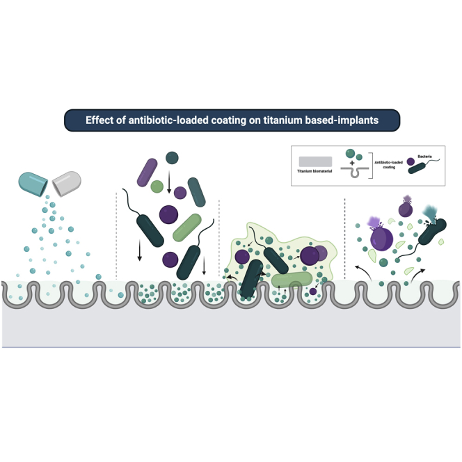
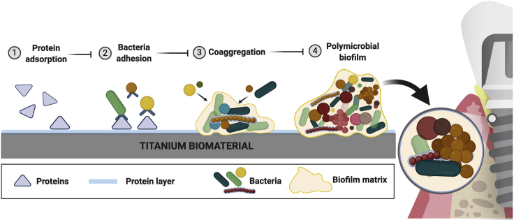
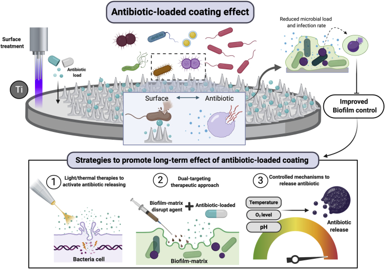
Implant devices have = proven a successful treatment modality in reconstructive surgeries. However, increasing rates of peri-implant diseases demand further examination of their pathogenesis. Polymicrobial biofilm formation on titanium surfaces has been considered the main risk factor for inflammatory processes on tissues surrounding implant devices, which often lead to implant failure. To overcome microbial accumulation on titanium surfaces biofilm targeting strategies have been developed to modify the surface and incorporate antimicrobial coatings. Because antibiotics are widely used to treat polymicrobial infections, these agents have recently started to be incorporated on titanium surface. This review discusses the biofilm formation on titanium dental implants and key factors to be considered in therapeutic and preventative strategies. Moreover, a systematic review was conducted on coatings developed for titanium surfaces using different antibiotics. This review will also shed light on potential alternative strategies aiming to reduce microbial loads and control polymicrobial infection on implanted devices.

Keywords: Microbiofilms; Surface Science.


Figures

None
Graphical abstract
Figure 1
Figure 1
Venn diagram of proteins adsorbed on titanium (Ti), enamel, and dentine surfaces from saliva (S) and plasma (P) Surfaces were exposed to human stimulated saliva or human plasma for 2 h at 35°C in an orbital shaker (70 rpm) and evaluated by liquid chromatography coupled with tandem mass spectrometry (LC–MS/MS). Numbers in parentheses are total proteins identified in each surface. Although there are shared proteins among the surfaces and in the different fluids (saliva and plasma), all groups/surfaces showed unique proteins adsorbed, showing the effect of chemical and physical properties of substrate to modulate protein adsorption.
Figure 2
Figure 2
Steps of biofilm formation on titanium biomaterial Dental implant surfaces made of titanium biomaterial provide the substrate for polymicrobial biofilm formation in the oral environment. Titanium surface is immediately coated by proteins from saliva (supra-mucosal segment) and plasma (sub-mucosal segment) after implant insertion. (1) Protein adsorption on the surface forms a layer with a composition directly affected by the chemical and physical properties of the surface. This layer is the main mediator of microbial adhesion through adhesin-receptor interactions. (2) Initial colonizers, mainly Streptococcus species, adhere on the surface binding to the protein layer. (3) Subsequently, co-aggregation processes and interaction between different species promote biofilm accumulation. (4) This synergistic interaction among organisms continues to contribute to the biofilm structure. These microbial communities are gradually embedded in the extracellular matrix, formed mainly by exopolysaccharides, eDNA, and proteins. This biofilm environment (structure) enhances the microbial interaction and cooperation, antimicrobial resistance, and nutrients/biomolecule retention-diffusion (created by BioRender®).
Figure 3
Figure 3
Candida and Streptococcus mixed-biofilms on titanium surface (A and B) Twenty-four-hour mixed biofilm stained by immuno-FISH of Candida albicans (green) and Streptococcus oralis (stained in blue on A and B and in red on C) growing on (A) titanium surface and (B) polystyrene surface. Bacterial extracellular matrix is stained with Alexa Fluor 647-labeled dextran conjugate probe (red) in figure (B). Images suggest that biofilm growth is modulated by the type of surface. (C) C. albicans (green) and S. oralis (red) interaction on biofilm growing on the polystyrene surface. (D) Dual species biofilms growing on the organotypic mucosal construct, which can cause epithelial barrier breach (H&E stain). (E) Titanium-mucosal interface biofilm model. Biofilm formed on titanium surface suspended 0.5 to 1 mm above the in vitro organotypic mucosal construct surface. Reprinted (adapted) from refs (Souza et al., 2020c, Souza et al., 2020d); Copyright (2020), with permission from American Society for Microbiology and Springer Nature. Figure E created by BioRender®.
Figure 4
Figure 4
Schematic representation of the “ecological plaque hypothesis” in relation to peri-implant disease, adapted from Marsh et al. (2011) and Rosier et al. (2018) Increased biofilm accumulation on implant surface triggers an inflammatory process that changes the environment leading to microbiological shift and disease progression, as shown by red boxes. Other factors can also favor the microbiological shift on biofilms growing on titanium surfaces, such as carbohydrate (sucrose exposure). However, some factors can control biofilm accumulation and inflammatory response, shown in blue boxes, such as surgical and antimicrobial intervention and host-response.
Figure 5
Figure 5
Schematic representation of the in situ model used to form biofilm on titanium surface using palatal appliances Sucrose is used to promote biofilm accumulation and extracellular matrix formation. Transmission electron microscopy showing biofilms formed in situ on Ti surface and exposed to Ti particles treatment. Red arrows showing Ti particles agglomerated and precipitated Ti ions on extracellular sites. Reprinted (adapted) from Ref. (Souza et al., 2019a); Copyright (2020), with permission from John Wiley and Sons.
Figure 6
Figure 6
Diagram of the source and selection procedures, according to the PRISMA guidelines
Figure 7
Figure 7
Word clouds of the antibiotics used on titanium coating in the studies included The font size represents the frequency of antibiotics used in which bigger words mean more frequent antibiotics used.
Figure 8
Figure 8
Percentage of reduction in the bacterial load on different antibiotic-loaded coatings on titanium surface in relation to control groups according to the study results included in this systematic review Circles represent each study included in the review in which it was possible to calculate bacterial load reduction and to compare studies using the same antibiotic coating.
Figure 9
Figure 9
Schematic representation of the anti-biofilm activity of antibiotic-loaded coatings on titanium surface Antibiotic loaded on titanium material by surface treatment may have bacteriostatic or bactericidal effect upon microbial contact with the antibiotic incorporated on the surface or by contact with the drug slowly released in the environment. This end result is reduction in microbial load of polymicrobial biofilms and, consequently, infection rates. Although a short-term releasing has been related to antibiotic-loaded coatings on titanium, some strategies (bottom panel) may promote drug releasing and antimicrobial effect, such as light therapies to activate the coating, dual-targeting therapies, and controlled mechanisms to release antibiotic (created by BioRender®).

References

    1. Abusleme L., Dupuy A.K., Dutzan N., Silva N., Burleson J.A., Strausbaugh L.D., Gamonal J., Diaz P.I. The subgingival microbiome in health and periodontitis and its relationship with community biomass and inflammation. ISME J. 2013;7:1016–1025. - PMC - PubMed
    1. Albrektsson T., Wennerberg A. Oral implant surfaces: Part 1–review focusing on topographic and chemical properties of different surfaces and in vivo responses to them. Int. J. Prosthodont. 2004;17:536–543. - PubMed
    1. Adams C.S., Antoci V., Jr., Harrison G., Patal P., Freeman T.A., Shapiro I.M., Parvizi J., Hickok N.J., Radin S., Ducheyne P. Controlled release of vancomycin from thin sol-gel films on implant surfaces successfully controls osteomyelitis. J. Orthop. Res. 2009;27:701–709. - PubMed
    1. Alt V., Kirchhof K., Seim F., Hrubesch I., Lips K.S., Mannel H., Domann E., Schnettler R. Rifampicin-fosfomycin coating for cementless endoprostheses: antimicrobial effects against methicillin-sensitive Staphylococcus aureus (MSSA) and methicillin-resistant Staphylococcus aureus (MRSA) Acta Biomater. 2014;10:4518–4524. - PubMed
    1. Antoci V., Jr., Adams C.S., Hickok N.J., Shapiro I.M., Parvizi J. Vancomycin bound to Ti rods reduces periprosthetic infection: preliminary study. Clin. Orthop. Relat. Res. 2007;461:88–95. - PubMed

LinkOut - more resources